

Review
doi: 10.1038/s41556-018-0133-0.
Epub 2018 Jun 27.
Mitochondrial dynamics in adaptive and maladaptive cellular stress responses
Affiliations
- PMID: 29950571
- PMCID: PMC6716149
- DOI: 10.1038/s41556-018-0133-0
Item in Clipboard
Review
Mitochondrial dynamics in adaptive and maladaptive cellular stress responses
Nat Cell Biol.
2018 Jul.
Abstract
Mitochondria sense and respond to many stressors and can support either cell survival or death through energy production and signaling pathways. Mitochondrial responses depend on fusion-fission dynamics that dilute and segregate damaged mitochondria. Mitochondrial motility and inter-organellar interactions, including with the endoplasmic reticulum, also function in cellular adaptation to stress. In this Review, we discuss how stressors influence these components, and how they contribute to the complex adaptive and pathological responses that lead to disease.
Conflict of interest statement
Authors declare that they do not have any competing interests.
Figures
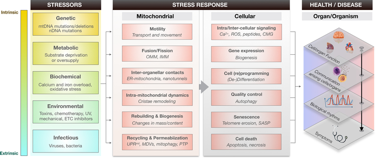
Stressors affecting mitochondria vary in nature and origin (left). Intrinsic stressors are those that arise from the molecular and biological components of the organism itself, such as DNA mutations and the product of chemical reactions. Stressors induce specific stress responses involving multiple facets of mitochondrial dynamics and key cellular processes (center). Components of the mitochondrial and cellular stress responses are not mutually exclusive, and also interact and influence each other (not depicted in the figure). Collectively, stress responses affect health and disease trajectories in a multi-level way by influencing inter-related domains of organ and organism function (right). Physiology and pathology can therefore manifest at each level, clinically at the level of symptoms and disorders (e.g., fatigue, ataxia, ophtalmoplegia), and sub-clinically in the disruption of biological rhythms (e.g., circadian oscillations, mitochondrial membrane potential oscillations), of cell-cell communication (e.g., production of mitochondria-derived metabolites, pro-inflammatory molecules and cytokines), or of organ systems (e.g., brain and cognitive function, cardiac contractility, hormone biosynthesis). Levels of function are interconnected. Abbreviations: mtDNA, mitochondrial DNA; nDNA, nuclear DNA; OMM, outer mitochondrial membrane; IMM, inner mitochondrial membrane; ER, endoplasmic reticulum; UPRmt, mitochondrial unfolded protein response; MDVs, mitochondria-derived vesicles; PTP, permeability transition pore; ROS, reactive oxygen species; CMG, circulating mitochondrial genome (also ccf-mtDNA); SASP, senescence-associated secretory profile; ETC, electron transport chain.
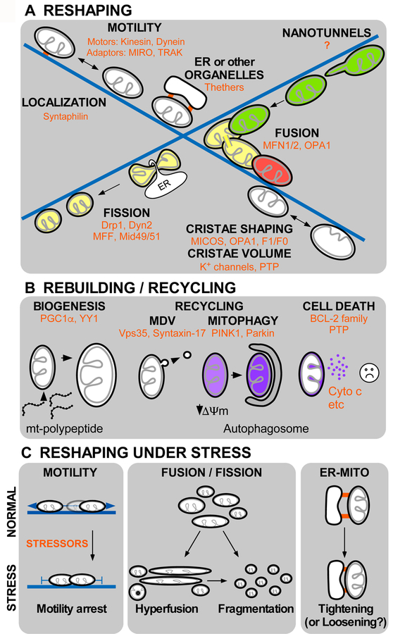
A. Reshaping, localization and motility of mitochondria (depicted by black OMM and gray IMM) along the microtubules (blue) supported by molecular motors (Kinesin and Dynein) and adaptors (MIRO and TRAK) facilitates the inter-organelle communication and physical tethering with the ER or other organelles. Mitochondrial fusion (green and red organelles merge to result yellow post-fusion content) occurs in association to microtubules and mediated by GTPase proteins located at the OMM (MFN1/2) and IMM (OPA1). Fission of mitochondria is also supported by association with the ER, and triggered by DRP1 and Dynamin2 GTPases. Recently described dynamic processes are mitochondrial nanotunnel formation that also depends on interaction with microtubules, intra-mitochondrial dynamics directed by MICOS (mitochondrial contact site and cristae organizing system), OPA1 and F1/F0 (ATP synthase) and matrix volume changes, depending on IMM K+ channels and the Permeability Transition Pore (PTP). B. Rebuilding and recycling processes, mitochondrial biogenesis involves expression of organelle-targeted proteins upon activation of transcriptional factors PGC1-α and YY1, and phospholipids biosynthesis. Recycling of mitochondria can be mediated by mitochondria derived vesicles (MDVs) regulated by Vps35, Syntaxin-17, and mitophagy, driven by PINK1 and Parkin. Mitochondria host cell death signaling pathways that control cytochrome c release to decide on cell survival or removal C. Mitochondrial reshaping under stress. Diverse stressors (red) trigger adaptive responses in mitochondrial reshaping processes. Stressors commonly cause mitochondrial motility arrest, hyperelongation and donut formation or total fragmentation. Under stress, ER-mitochondria contacts usually become tighter but loosening has also been documented.
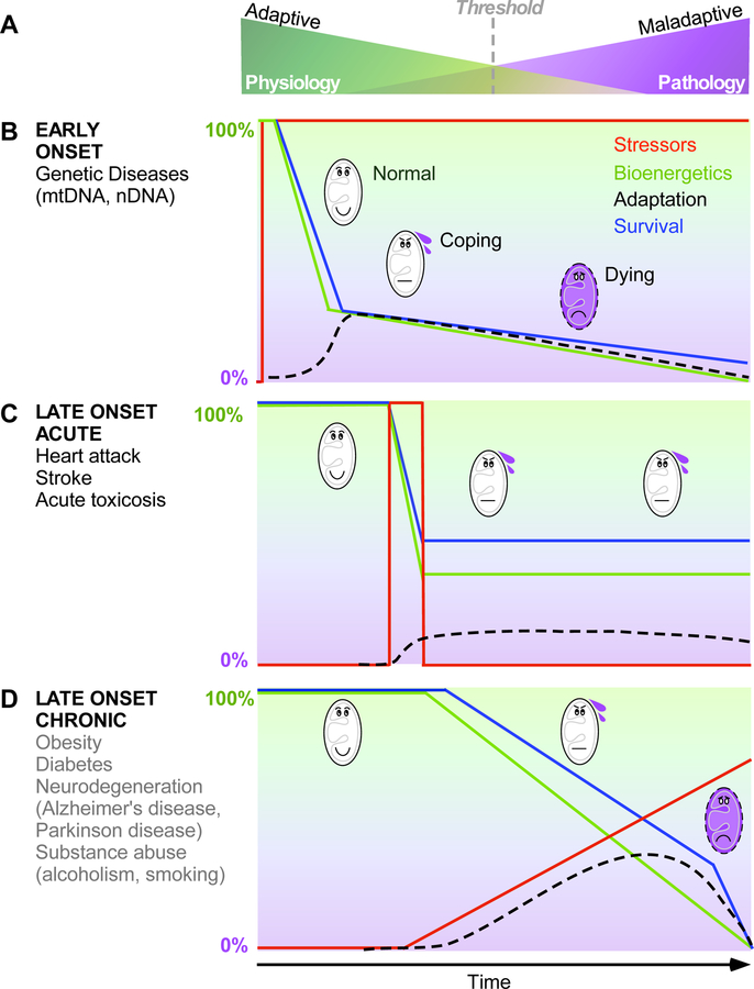
A. Biological systems stand in balance between adaptative and maladaptative states that determine physiological and pathological outcomes. The threshold between these states can vary between individuals, and over time. B-D. Three main types of stressors can be identified based on their onset and duration. Stressors refer to a singular perturbation with a time of onset and specific duration; Bioenergetics is the capacity to use OXPHOS to transform energetic substrates and oxygen into ΔΨm to generate ATP and perform work (e.g., Ca2+ uptake), illustrated here as the transition from green to purple; Adaptation reflects the activation of secondary processes (e.g., gene expression, mitochondrial biogenesis, increased contractility) that act to compensate for bioenergetic defects. Survival indicates the ability of cells and organs to sustain viability and normal functions. B. EARLY ONSET stressors are generally chronic in nature, such as inherited mtDNA and nDNA mutations that alter key components of mitochondrial dynamics and bioenergetics. Early onset chronic stressors may cause a substantial initial loss in bioenergetic capacity (i.e., fitness) and lead to progressive decline in survival. C. LATE ONSET ACUTE stressors are punctual, arising from chemical exposure, ischemia, or other reversible event. Late onset acute stressors generally induce a rapid and substantial loss of bioenergetic capacity and survival associated with an induction of compensatory adaptive processes that may remain elevated beyond the duration of the stressor, enabling the maintenance of sub-maximal but sufficient functional capacity. D. LATE ONSET CHRONIC stressors are those that also arise punctually later in life but remain active and often progress in intensity, such as metabolic dysregulation in diabetes, neurodegenerative processes, and toxic compound exposure from substance abuse. Late onset chronic stressors generally lead to progressive decline in bioenergetics and survival, the progression of which is reduced by compensatory adaptive mechanisms.
Comment in
-
Focusing on mitochondrial form and function.Nat Cell Biol. 2018 Jul;20(7):735. doi: 10.1038/s41556-018-0139-7. Nat Cell Biol. 2018. PMID: 29950569 No abstract available.
References
Publication types
MeSH terms
Grants and funding
LinkOut - more resources
Full Text Sources
Other Literature Sources
