Review
. 2018 Oct;15(10):631-647.
doi: 10.1038/s41569-018-0046-4.

The epicardium as a hub for heart regeneration

Affiliations
Review

The epicardium as a hub for heart regeneration

Jingli Cao et al. Nat Rev Cardiol. 2018 Oct.

Abstract

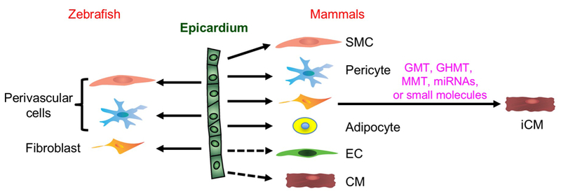
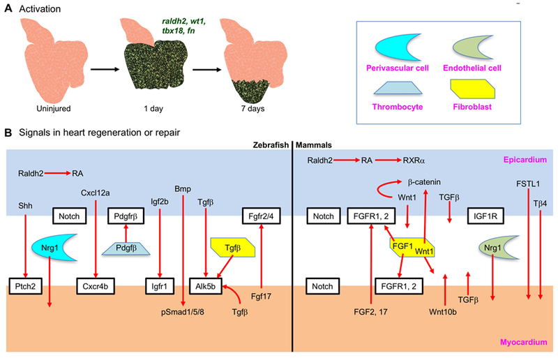
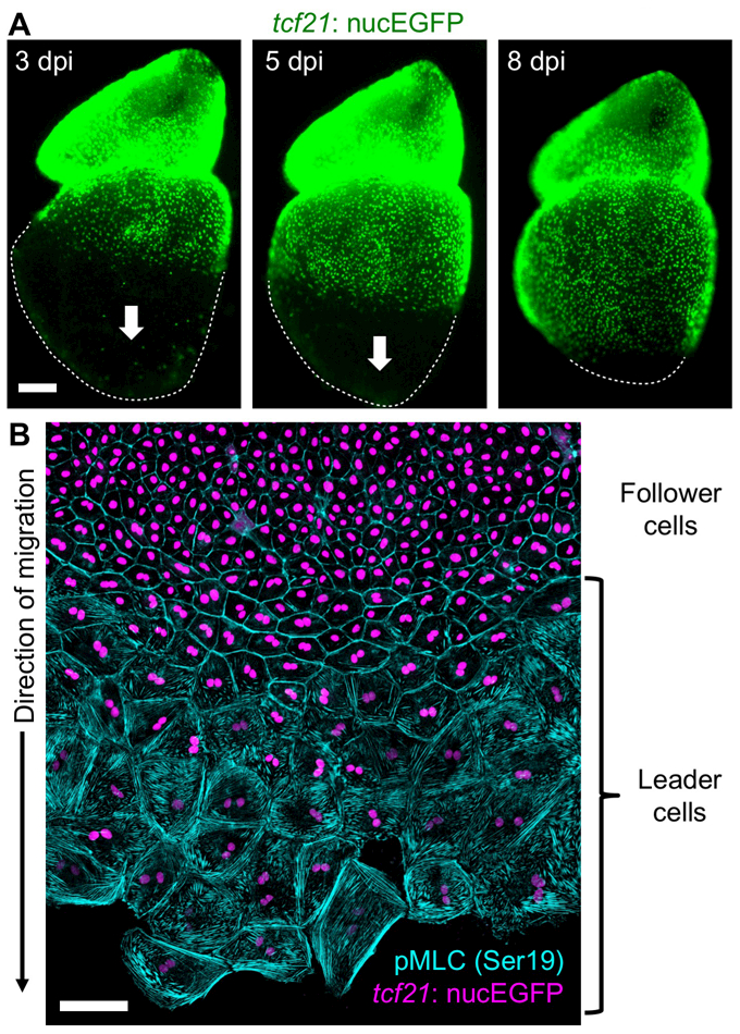
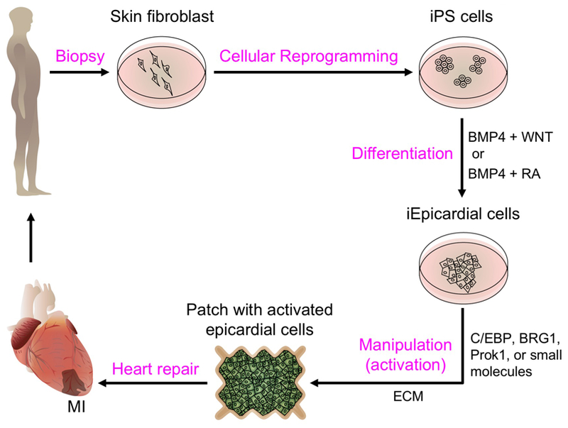
After decades of directed research, no effective regenerative therapy is currently available to repair the injured human heart. The epicardium, a layer of mesothelial tissue that envelops the heart in all vertebrates, has emerged as a new player in cardiac repair and regeneration. The epicardium is essential for muscle regeneration in the zebrafish model of innate heart regeneration, and the epicardium also participates in fibrotic responses in mammalian hearts. This structure serves as a source of crucial cells, such as vascular smooth muscle cells, pericytes, and fibroblasts, during heart development and repair. The epicardium also secretes factors that are essential for proliferation and survival of cardiomyocytes. In this Review, we describe recent advances in our understanding of the biology of the epicardium and the effect of these findings on the candidacy of this structure as a therapeutic target for heart repair and regeneration.


Conflict of interest statement

COMPETING INTERESTS

The authors declare they have no conflicts of interest.

Figures

Figure 1.
Figure 1.. Cellular contributions of epicardial cells during zebrafish heart regeneration and mammalian heart repair.
CM, cardiomyocyte; iCM, induced cardiomyocyte through reprogramming; SMC, smooth muscle cell; EC, endothelial cell; GMT, Gata4 + Mef2c + Tbx5; GHMT, GMT + Hand2; MMT, Mef2c + Myocd + Tbx5.
Figure 2.
Figure 2.. Epicardial signals in heart repair and regeneration.
(A) Organ-wide activation of zebrafish epicardial cells one day after resection of the ventricular apex. Activation (induction of embryonic epicardial markers such as raldh2, wt1, tbx18 and fn) is restricted to the injury site by 7 days after injury. Green color represents the re-expression of embryonic genes. (B) Proposed epicardium-myocardium signals in zebrafish heart regeneration and mammalian heart repair.
Figure 3.
Figure 3.. Ex vivo epicardial regeneration.
(A) Whole-mount images of explanted zebrafish heart showing epicardial regeneration along the ventricular surface in a base-to-apex direction (arrows). tcf21:nucEGFP visualizes epicardial cell nuclei (green). dpi, days post Mtz incubation. (B) The epicardium generates a leading edge of large, multinucleate (leader, bottom) cells during migration ex vivo. Trailing (follower, top) cells are small and mononucleate. Epicardial nuclei are indicated in violet (tcf21:nucEGFP) and phosphorylated myosin light chain 2 (Ser19), an indicator of mechanical tension, is cyan.
Figure 4.
Figure 4.. An epicardium-derived strategy for heart repair.
Skin fibroblasts from the MI patient could be reprogrammed into induced pluripotent stem cells (iPS cells) and further differentiated into induced epicardial cells (iEpicardial cells) by defined factors (see section 6.3). iEpicardial cells could be activated through treatment with small molecules or pro-regenerative factors (sections 4 and 6.1), and engineered with ECM components (section 4.2) into a patch (section 6.2) to be applied to the MI.

References

    1. Acharya A, Baek ST, Huang G, Eskiocak B, Goetsch S, Sung CY, Banfi S, Sauer MF, Olsen GS, Duffield JS, et al. (2012). The bHLH transcription factor Tcf21 is required for lineage-specific EMT of cardiac fibroblast progenitors. Development 139, 2139–2149. - PMC - PubMed
    1. Ali SR, Ranjbarvaziri S, Talkhabi M, Zhao P, Subat A, Hojjat A, Kamran P, Muller AM, Volz KS, Tang Z, et al. (2014). Developmental heterogeneity of cardiac fibroblasts does not predict pathological proliferation and activation. Circ Res 115, 625–635. - PubMed
    1. Athanasiadis EI, Botthof JG, Andres H, Ferreira L, Lio P, and Cvejic A (2017). Single-cell RNA-sequencing uncovers transcriptional states and fate decisions in haematopoiesis. Nat Commun 8, 2045. - PMC - PubMed
    1. Balmer GM, Bollini S, Dube KN, Martinez-Barbera JP, Williams O, and Riley PR (2014). Dynamic haematopoietic cell contribution to the developing and adult epicardium. Nat Commun 5, 4054. - PMC - PubMed
    1. Bao X, Lian X, Hacker TA, Schmuck EG, Qian T, Bhute VJ, Han T, Shi M, Drowley L, Plowright A, et al. (2016). Long-term self-renewing human epicardial cells generated from pluripotent stem cells under defined xeno-free conditions. Nat Biomed Eng 1. - PMC - PubMed

Publication types

MeSH terms