

IL-6, PF-4, sCD40 L, and homocysteine are associated with the radiological progression of cerebral small-vessel disease: a 2-year follow-up study
- PMID: 29950823
- PMCID: PMC6016008
- DOI: 10.2147/CIA.S166773
IL-6, PF-4, sCD40 L, and homocysteine are associated with the radiological progression of cerebral small-vessel disease: a 2-year follow-up study
Abstract
Background: Endothelial dysfunction (ED) is involved in the pathogenesis of cerebral small vessel disease (SVD), however, it is not clear if specific biomarkers related to ED are associated with radiological progression of SVD.
Methods: A single-center, prospective cohort study was conducted among consecutive, adult patients with SVD. Logistic regression was used to analyze the association of each baseline biomarker (highest vs lowest tertile) and the MRI radiological outcome after 2 years. The mean Z-score for vascular inflammation (VI) combined soluble intercellular cell adhesion molecule-1 (sICAM-1), soluble platelet selectin (sP-selectin), CD40 ligand (sCD40 L), platelet factor-4 (PF-4) and homocysteine; Z-score for systemic inflammation (SI) combined high-sensitivity C-reactive protein (hsCRP), interleukin-1α and -6 (IL-1α and IL-6, respectively) and tumor necrosis factor-α (TNF-α).
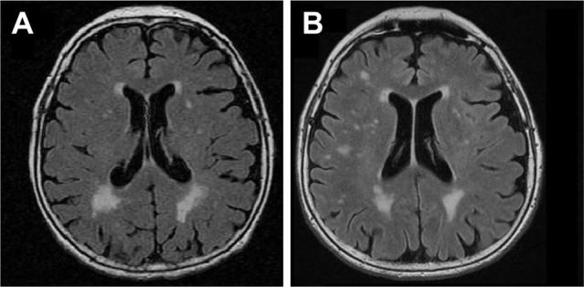
Results: The study group comprised 123 patients (age, mean±SD: 72.2±8 years, 49% females), with lacunar stroke (n=49), vascular dementia (n=48), and vascular parkinsonism (n=26). Moreover, 34.9% patients experienced radiological progression, 43% had progression of isolated white matter lesions (WMLs), 23.2% had new lacunes, and 34.8% had both WMLs progression and new lacunes. After adjustment for confounders (age, sex, blood pressure, MRI lesions load), the PF-4 (OR; 95% CI 5.5; 1.5-21), sCD40L (4.6; 1.1-18.6), IL-6 (7.4; 1.48-37), Z-score for VI (4.5; 1.1-18.6), and, marginally, homocysteine (4.1; 0.99-17) were associated with the risk of any radiological progression; further, homocysteine (2.4; 1.4-14), Z-score for SI (2.1; 1.2-14) and, marginally, IL-6 (6.0; 0.95 -38) were related to the development of new lacunes; PF-4 (7.9; 1.6-38) and, marginally, the Z-score for VI (4.2; 0.9-19.5) were correlated with the risk of WMLs progression. Additional adjustment for clinical SVD manifestations did not significantly alter the results.
Conclusion: The data supports the concept that ED modulates the radiological progression of SVD and WMLs and lacunes are associated with different inflammatory markers.
Keywords: IL-6; PF-4; cerebral small-vessel disease; homocysteine; radiological progression; sCD40 L.
Conflict of interest statement
Disclosure The authors report no conflicts of interest in this work.
Figures
Similar articles
-
Association between hemostatic markers, serum lipid fractions and progression of cerebral small vessel disease: A 2-year follow-up study.Neurol Neurochir Pol. 2018 Jan-Feb;52(1):54-63. doi: 10.1016/j.pjnns.2017.11.005. Epub 2017 Nov 14. Neurol Neurochir Pol. 2018. PMID: 29173807
-
Inflammatory biomarkers and cerebral small vessel disease: a community-based cohort study.Stroke Vasc Neurol. 2022 Aug;7(4):302-309. doi: 10.1136/svn-2021-001102. Epub 2022 Mar 8. Stroke Vasc Neurol. 2022. PMID: 35260438 Free PMC article.
-
IL-1α and IL-6 predict vascular events or death in patients with cerebral small vessel disease-Data from the SHEF-CSVD study.Adv Med Sci. 2019 Sep;64(2):258-266. doi: 10.1016/j.advms.2019.02.003. Epub 2019 Mar 4. Adv Med Sci. 2019. PMID: 30844663
-
Inflammation and cerebral small vessel disease: A systematic review.Ageing Res Rev. 2019 Aug;53:100916. doi: 10.1016/j.arr.2019.100916. Epub 2019 Jun 10. Ageing Res Rev. 2019. PMID: 31181331
-
Diffusion magnetic resonance imaging in cerebral small vessel disease.Rev Neurol (Paris). 2017 Apr;173(4):201-210. doi: 10.1016/j.neurol.2017.03.005. Epub 2017 Apr 6. Rev Neurol (Paris). 2017. PMID: 28392060 Review.
Cited by
-
Blood biomarkers of vascular dysfunction in small vessel disease progression: Insights from a longitudinal neuroimaging study.Alzheimers Dement. 2025 Apr;21(4):e70152. doi: 10.1002/alz.70152. Alzheimers Dement. 2025. PMID: 40275856 Free PMC article.
-
Emerging Biomarkers in Vascular Cognitive Impairment and Dementia: From Pathophysiological Pathways to Clinical Application.Int J Mol Sci. 2019 Jun 8;20(11):2812. doi: 10.3390/ijms20112812. Int J Mol Sci. 2019. PMID: 31181792 Free PMC article. Review.
-
Small Vessel Disease-Related Dementia: An Invalid Neurovascular Coupling?Int J Mol Sci. 2020 Feb 7;21(3):1095. doi: 10.3390/ijms21031095. Int J Mol Sci. 2020. PMID: 32046035 Free PMC article. Review.
-
Immune regulation and blood-brain barrier permeability in cerebral small vessel disease: study protocol of the INflammation and Small Vessel Disease (INSVD) study - a multicentre prospective cohort study.BMJ Open. 2024 Feb 26;14(2):e084303. doi: 10.1136/bmjopen-2024-084303. BMJ Open. 2024. PMID: 38413153 Free PMC article.
-
Serum Neurofilament Light Chain Is Associated with Incident Lacunes in Progressive Cerebral Small Vessel Disease.J Stroke. 2020 Sep;22(3):369-376. doi: 10.5853/jos.2019.02845. Epub 2020 Sep 29. J Stroke. 2020. PMID: 33053952 Free PMC article.
References
-
- Pantoni L. Cerebral small vessel disease: from pathogenesis and clinical characteristics to therapeutic challenges. Lancet Neurol. 2010;9(7):689–701. - PubMed
-
- Staszewski J, Piusińska-Macoch R, Brodacki B, Skrobowska E, Stępień A. Association between hemostatic markers, serum lipid fractions and progression of cerebral small vessel disease: a 2-year follow-up study. Neurol Neurochir Pol. 2018;52(1):54–63. - PubMed
MeSH terms
Substances
LinkOut - more resources
Full Text Sources
Other Literature Sources
Research Materials
Miscellaneous
