. 2018 May 21:2018:6463807.
doi: 10.1155/2018/6463807. eCollection 2018.

Geranylgeraniol Prevents Statin-Dependent Myotoxicity in C2C12 Muscle Cells through RAP1 GTPase Prenylation and Cytoprotective Autophagy

Affiliations

Geranylgeraniol Prevents Statin-Dependent Myotoxicity in C2C12 Muscle Cells through RAP1 GTPase Prenylation and Cytoprotective Autophagy

Anna Jaśkiewicz et al. Oxid Med Cell Longev. .

Abstract

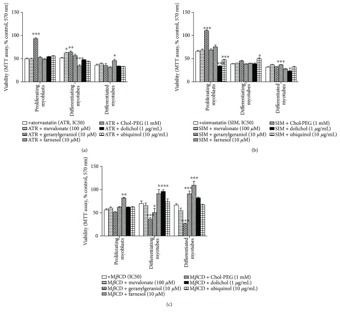
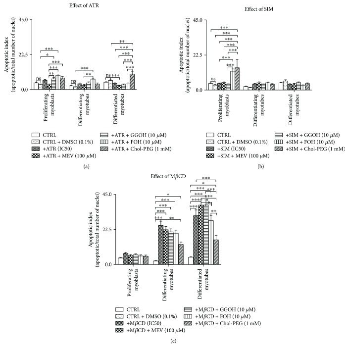
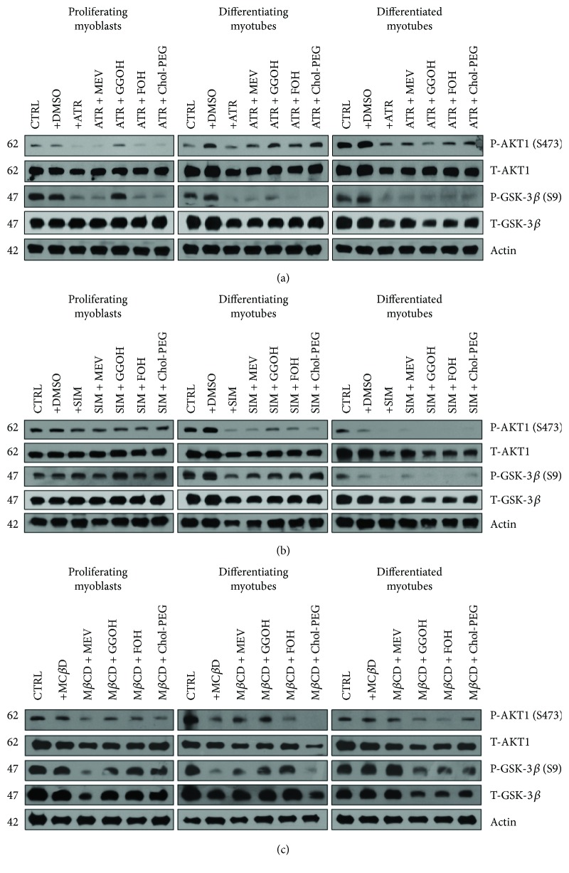
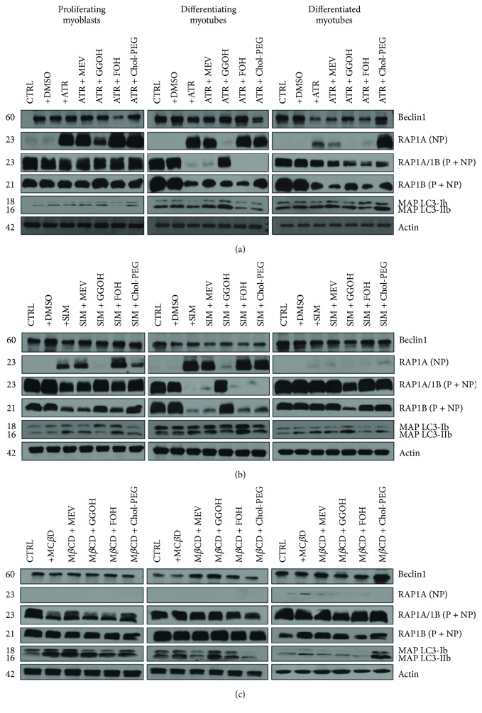
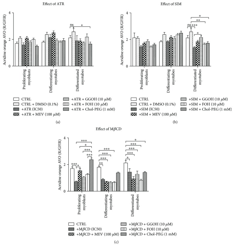
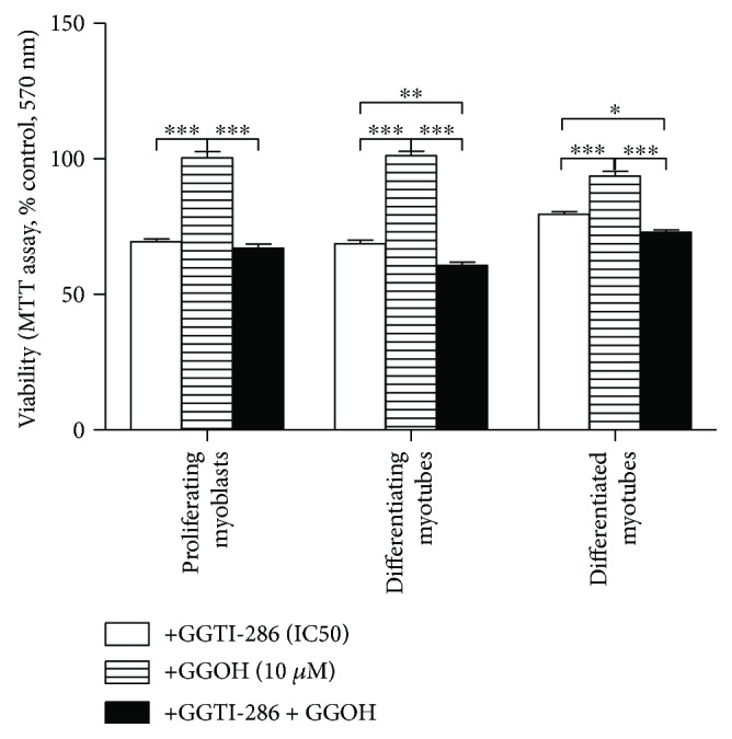
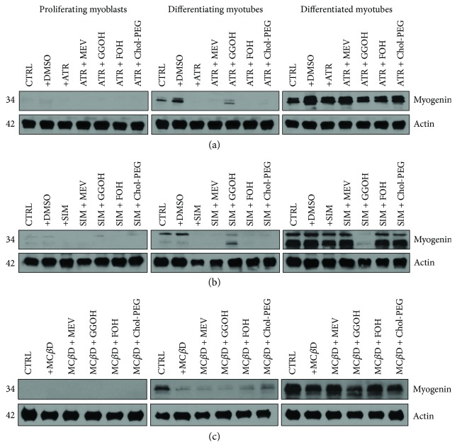
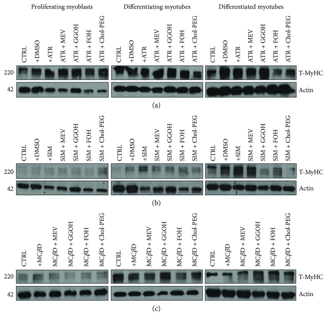
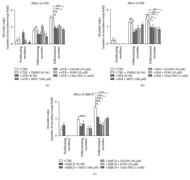
The present study investigated the cytotoxic effects of statins (atorvastatin (ATR) and simvastatin (SIM), resp.) and methyl-beta-cyclodextrin (MβCD), at their respective IC50 concentrations, on muscle regeneration in the in vitro model of murine C2C12 myoblasts. Cotreatment with mevalonate (MEV), farnesol (FOH), geranylgeraniol (GGOH), or water-soluble cholesterol (Chol-PEG) was employed to determine whether the statin-dependent myotoxicity resulted from the lower cholesterol levels or the attenuated synthesis of intermediates of mevalonate pathway. Our findings demonstrated that while GGOH fully reverted the statin-mediated cell viability in proliferating myoblasts, Chol-PEG exclusively rescued MβCD-induced toxicity in myocytes. Statins caused loss of prenylated RAP1, whereas the GGOH-dependent positive effect was accompanied by loss of nonprenylated RAP1. Geranylgeranyltransferases are essential for muscle cell survival as inhibition with GGTI-286 could not be reversed by GGOH cotreatment. The increase in cell viability correlated with elevated AKT 1(S463) and GSK-3β(S9) phosphorylations. Slight increase in the levels of autophagy markers (Beclin 1, MAP LC-3IIb) was found in response to GGOH cotreatment. Autophagy rose time-dependently during myogenesis and was inhibited by statins and MβCD. Statins and MβCD also suppressed myogenesis and neither nonsterol isoprenoids nor Chol-PEG could reverse this effect. These results point to GGOH as the principal target of statin-dependent myotoxicity, whereas plasma membrane cholesterol deposit is ultimately essential to restore viability of MβCD-treated myocytes. Overall, this study unveils for the first time a link found between the GGOH- and Chol-PEG-dependent reversal of statin- or MβCD-mediated myotoxicity and cytoprotective autophagy, respectively.


Figures

Figure 1
Figure 1
Effect of nonsterol isoprenoids and soluble cholesterol treatments on C2C12 muscle cell viability. Nonsterol isoprenoids and soluble cholesterol differentially rescue C2C12 myoblasts from statin- or MβCD-reduced (IC50) cell viability. C2C12 myoblasts were exposed for 24, 72, or 120 h to statins or MβCD (IC50), (day 1—proliferating myoblasts, day 3—differentiating myotubes, and day 5—differentiated myotubes). ATR or SIM was administered in the listed concentration at each day of differentiation: ATR: day 1—76 μM, day 3—46 μM, and day 5—36 μM; SIM: day 1—87 μM, day 3—6 μM, and day 5—3 μM. MβCD was added to give the final concentration: day 1—2.7 mM, day 3—1.9 mM, and day 5—1.1 mM or vehicle control (0.1% DMSO or 2% HS DMEM) without or with selected mevalonate pathway intermediate (mevalonate 100 μM, geranylgeraniol 10 μM, farnesol 10 μM, Chol-PEG 1 mM, dolichol 1 μg/mL, and ubiquinol 10 μg/mL). (a) Geranylgeraniol (GGOH, 10 μM) in proliferating myoblasts and differentiating myotubes, farnesol (FOH, 10 μM) in differentiating myotubes, and Chol-PEG in differentiated myotubes inhibited ATR-dependent drop in cell viability. (b) Geranylgeraniol (GGOH, 10 μM) in proliferating myoblasts, ubiquinol in differentiating myotubes, and farnesol in differentiated myotubes inhibited SIM-dependent drop in cell viability. (c) Soluble cholesterol (Chol-PEG, 1 mM) in proliferating myoblasts, differentiating myotubes, and differentiated myotubes; dolichol (DOH, 1 μg/mL) in differentiating myotubes; and mevalonate (MEV, 100 μM) in differentiated myotubes inhibited MβCD-dependent drop in cell viability. Two-way ANOVA test [time (proliferating myoblasts, differentiating myotubes, differentiated myotubes)] amounted to F(2,187) = 201.73, p < 0.0001 for ATR; F(2,227) = 665.22, p < 0.0001 for SIM; F(2,371) = 8.58, p < 0.0002 for MβCD. Treatment: F(6,187) = 38.96, p < 0.0001 (ATR, ATR + MEV, ATR + GGOH, ATR + FOH, and ATR + Chol-PEG); F(6,227) = 56.33, p < 0.0001 (SIM, SIM + MEV, SIM + GGOH, SIM + FOH, and SIM + Chol-PEG); F(6,371) = 58.43, p < 0.0001 (MβCD, MβCD + MEV, MβCD + GGOH, MβCD + FOH, and MβCD + Chol-PEG). Interaction: F(12,187) = 30.26, p < 0.0001 for ATR; F(12,227) = 49.40, p < 0.0001 for SIM; F(12,227) = 11.29, p < 0.0001 for MβCD. Error bars = SEM and p < 0.05, ∗∗p < 0.01, and ∗∗∗p < 0.001 for comparison with nontreated control cells. Results are means ± SEM of three independent experiments.
Figure 2
Figure 2
Effect of nonsterol isoprenoids and soluble cholesterol treatments on apoptotic index (AI) in C2C12 myoblasts affected by statins or MβCD. C2C12 myoblasts were exposed for 24, 72, or 120 h to statins or MβCD (IC50), (day 1—proliferating myoblasts, day 3—differentiating myotubes, and day 5—differentiated myotubes). ATR or SIM was administered in the listed concentration at each day of differentiation: ATR: day 1—76 μM, day 3—46 μM, and day 5—36 μM; SIM: day 1—87 μM, day 3—6 μM, and day 5—3 μM. MβCD was added to give the final concentration: day 1—2.7 mM, day 3—1.9 mM, and day 5—1.1 mM or vehicle control (0.1% DMSO or 2% HS DMEM) without or with selected mevalonate pathway intermediate (mevalonate 100 μM, geranylgeraniol 10 μM, farnesol 10 μM, and Chol-PEG 1 mM). Next, cells were subjected to vital staining with HO33342 (see Materials and Methods). Apoptotic index (AI) was calculated as percent value of apoptotic nuclei/total number of nuclei in at least 10 replicates for each treatment and nontreated controls. Two-way ANOVA test for AI followed by Bonferroni's multiple comparisons was employed to analyze the data. The results of [time (proliferating myoblasts, differentiating myotubes, differentiated myotubes)] amounted to F(2,189) = 17.46, p < 0.0001 for ATR; F(2,189) = 12.17, p < 0.0001 for SIM; F(2,162) = 142.3, p < 0.0001 for MβCD. Treatment: ATR, ATR + MEV, ATR + GGOH, ATR + FOH, and ATR + Chol-PEG (F(6,189) = 15.52, p < 0.0001), SIM, SIM + MEV, SIM + GGOH, SIM + FOH, and SIM + Chol-PEG (F(6,189) = 4.712, p = 0.0002), MβCD, MβCD + MEV, MβCD + GGOH, MβCD + FOH, and MβCD + Chol-PEG (F(5,162) = 37.52, p < 0.0001). Interaction: F(12,189) = 7.629, p < 0.0001 for ATR; F(12,189) = 5.111, p < 0.0001 for SIM; F(10,162) = 9.708, p < 0.0001 for MβCD. Error bars = SEM and p < 0.05, ∗∗p < 0.01, ∗∗∗p < 0.001 for comparison between the means. Results are means of three independent experiments.
Figure 3
Figure 3
Effect of nonsterol isoprenoids and soluble cholesterol treatments on AKT/GSK-3β in C2C12 myoblasts affected by statins or MβCD.
Figure 4
Figure 4
Effect of nonsterol isoprenoids and soluble cholesterol treatments on Beclin 1 and MAP LC3-Ib/MAP LC3-IIb and RAP GTPase in C2C12 myoblasts affected by statins or MβCD.
Figure 5
Figure 5
Effect of nonsterol isoprenoids and soluble cholesterol treatments on acidic vacuolar organelles (AVO) as red to green fluorescence intensity ratio (R/GFIR) in C2C12 myoblasts affected by statins or MβCD. C2C12 myoblasts were exposed for 24, 72, or 120 h to statins or MβCD (IC50) (day 1—proliferating myoblasts, day 3—differentiating myotubes, and day 5—differentiated myotubes). ATR or SIM was administered in the listed concentration at each day of differentiation: ATR: day 1—76 μM, day 3—46 μM, and day 5—36 μM; SIM: day 1—87 μM, day 3—6 μM, and day 5–3 μM. MβCD was added to give the final concentration: day 1—2.7 mM, day 3—1.9 mM, and day 5—1.1 mM or vehicle control (0.1% DMSO or 2% HS DMEM) without or with selected mevalonate pathway intermediate (mevalonate 100 μM, geranylgeraniol 10 μM, farnesol 10 μM, Chol-PEG 1 mM, dolichol 1 μg/mL, and ubiquinol 10 μg/mL). Next, cells were subjected to vital staining with acridine orange (see Materials and Methods). Red to green fluorescence intensity ratio (R/GFIR) was calculated in at least 10 replicates for each treatment and nontreated controls. Two-way ANOVA test for R/GFIR followed by Bonferroni's multiple comparisons was employed to analyze the data. The results of [time (proliferating myoblasts, differentiating myotubes, differentiated myotubes)] amounted to F(2,191) = 3.774, p = 0.0247 for ATR; F(2,194) = 11.21, p < 0.0001 for SIM; and F(2,189) = 11.78, p < 0.0001 for MβCD. Treatment: ATR, ATR + MEV, ATR + GGOH, ATR + FOH, and ATR + Chol-PEG (F(6,191) = 5.084, p < 0.0001); SIM, SIM + MEV, SIM + GGOH, SIM + FOH, and SIM + Chol-PEG (F(6,194) = 6.814, p < 0.0001); MβCD, MβCD + MEV, MβCD + GGOH, MβCD + FOH, and MβCD + Chol-PEG (F(5,189) = 23.42, p < 0.0001). Interaction: F(12,191) = 1.450, p = 0.1464 for ATR; F(12,194) = 2.604, p = 0.0031 for SIM; F(10,189) = 5.532, p < 0.0001 for MβCD. Error bars = SEM and p < 0.05, ∗∗p < 0.01, and ∗∗∗p < 0.001 for comparison between the means. Results are means of three independent experiments.
Figure 6
Figure 6
Geranylgeraniol (GGOH) does not rescue C2C12 myoblasts from GGTI-286-induced (IC50) compromised cell viability. GGTI-286 was used as a specific inhibitor of protein geranylgeranyltransferases. C2C12 myoblasts were exposed for 24, 72, or 120 h to GGTI-286 (day 1—proliferating myoblasts, day 3—differentiating myotubes, and day 5—differentiated myotubes). GGTI-286 (IC50): day 1—25 μM, day 3—24 μM, and day 5—23 μM or vehicle control (0.1% DMSO or 2% HS DMEM) without or with GGOH. GGOH (10 μM) could not reverse the drop in cell viability induced by GGTI-286. Two-way ANOVA test [time (proliferating myoblasts, differentiating myotubes, differentiated myotubes)] amounted to F(2, 42) = 10.69, p = 0.0002; treatment: GGTI-286, GGOH, and GGTI-286 + GGOH: F(2, 21) = 346.8, p < 0.0001; interaction: F(4, 42) = 18.73, p < 0.0001, followed by Bonferroni's multiple comparisons employed to analyze the data. Error bars = SEM and p < 0.05, ∗∗p < 0.01, and ∗∗∗p < 0.001 for comparison between the means. Results are means of three independent experiments.
Figure 7
Figure 7
Effect of nonsterol isoprenoids and soluble cholesterol treatments on myogenin in C2C12 myoblasts affected by statins or MβCD.
Figure 8
Figure 8
Effect of nonsterol isoprenoids and soluble cholesterol treatments on total myosin heavy chain (T-MyHC) amount in C2C12 myoblasts affected by statins or MβCD.
Figure 9
Figure 9
Effect of nonsterol isoprenoids and soluble cholesterol treatments on myotube index (MI) in C2C12 myoblasts affected by statins or MβCD. C2C12 myoblasts were exposed for 24, 72, or 120 h to statins or MβCD (IC50) (day 1—proliferating myoblasts, day 3—differentiating myotubes, and day 5—differentiated myotubes). ATR or SIM was administered in the listed concentration at each day of differentiation: ATR: day 1—76 μM, day 3—46 μM, and day 5—36 μM; SIM: day 1—87 μM, day 3—6 μM, and day 5—3 μM. MβCD was added to give the final concentration: day 1—2.7 mM, day 3—1.9 mM, and day 5–1.1 mM or vehicle control (0.1% DMSO or 2% HS DMEM) without or with selected mevalonate pathway intermediate (mevalonate 100 μM, geranylgeraniol 10 μM, farnesol 10 μM, Chol-PEG 1 mM, dolichol 1 μg/mL, and ubiquinol 10 μg/mL). Next, cells were subjected to immunofluorescence with HO33342 to counterstain nuclei (see Materials and Methods). The myotube index was determined as the ratio of the nuclear number in myotubes (C2C12 cells with three or more nuclei) to the total number of nuclei multiplied by 100%). (a) Atorvastatin (ATR, IC50) did not affect myotube index (MI) at days 1 and 3. However, it significantly diminished fraction of myonuclei in myotubes at day 5. Neither of added nonsterol isoprenoids nor cholesterol treatment could influence ATR effect. (b) Simvastatin (SIM, IC50) did not change MI at day 1, but it significantly lessened myotube representation at days 3 and 5. Nonsterol isoprenoids and cholesterol reversed SIM effect at day 3 but not at day 5. (c) Methyl-beta-cyclodextrin (MβCD, IC50) could not effect MI at day 1, but it significantly decreased percentage of myotubes at days 3 and 5 without any effect of nonsterol isoprenoids or cholesterol. Two-way ANOVA test for MI followed by Bonferroni's multiple comparisons was employed to analyze the data. The results of [time (proliferating myoblasts, differentiating myotubes, differentiated myotubes)] amounted to F(2,337) = 80.99, p < 0.0001 for ATR; F(2,352) = 98.91, p < 0.0001 for SIM; and F(2,266) = 59.00, p < 0.0001 for MβCD. Treatment: ATR, ATR + MEV, ATR + GGOH, ATR + FOH, and ATR + Chol-PEG (F(6,337) = 5.982, p < 0.0001); SIM, SIM + MEV, SIM + GGOH, SIM + FOH, and SIM + Chol-PEG (F(6,352) = 6.370, p < 0.0001); MβCD, MβCD + MEV, MβCD + GGOH, MβCD + FOH, and MβCD + Chol-PEG (F(5,266) = 14.77, p < 0.0001). Interaction: F(12,337) = 3.096, p = 0.0004 for ATR; F(12,352) = 0.8962, p = 0.5511 for SIM; F(10,266) = 2.560, p = 0.0057 for MβCD. Error bars = SEM and p < 0.05, ∗∗p < 0.01, and ∗∗∗p < 0.001 for comparison between the means. Results are means of three independent experiments.

References

    1. Endo A. The discovery and development of HMG-CoA reductase inhibitors. Journal of Lipid Research. 1992;33(11):1569–1582. - PubMed
    1. Liao J. K. Clinical implications for statin pleiotropy. Current Opinion in Lipidology. 2005;16(6):624–629. doi: 10.1097/01.mol.0000191913.16321.60. - DOI - PubMed
    1. Liao J. K., Laufs U. Pleiotropic effects of statins. Annual Review of Pharmacology and Toxicology. 2005;45(1):89–118. doi: 10.1146/annurev.pharmtox.45.120403.095748. - DOI - PMC - PubMed
    1. Liao J. K. Isoprenoids as mediators of the biological effects of statins. Journal of Clinical Investigation. 2002;110(3):285–288. doi: 10.1172/JCI0216421. - DOI - PMC - PubMed
    1. Rosenson R. S. Current overview of statin-induced myopathy. The American Journal of Medicine. 2004;116(6):408–416. doi: 10.1016/j.amjmed.2003.10.033. - DOI - PubMed

MeSH terms

LinkOut - more resources