. 2018 Jun 20:4:24.
doi: 10.1186/s40942-018-0127-x. eCollection 2018.

Dexamethasone implant in silicone oil: in vitro behavior

Affiliations

Dexamethasone implant in silicone oil: in vitro behavior

Erick Omar Flores-Villalobos et al. Int J Retina Vitreous. .

Abstract

Background: To determine the effect of the silicone on the dexamethasone intravitreal implant.

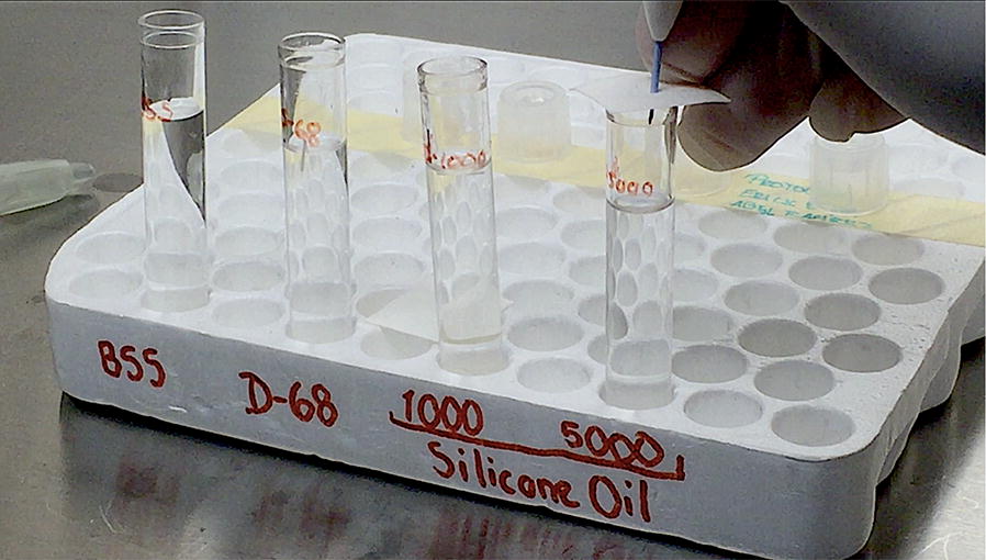
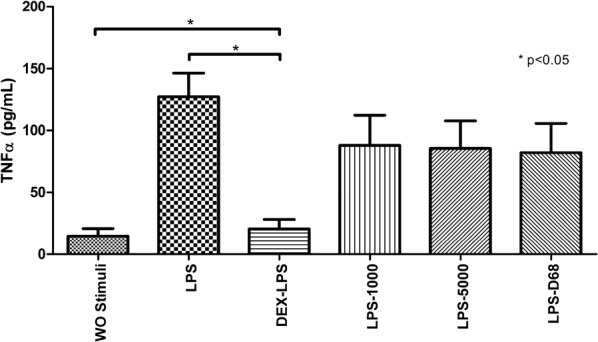
Methods: Basic, experimental, prospective and transversal study performed at the hospital "Nuestra Señora de la Luz" in Mexico City. One dexamethasone implant was placed in a test tube with 4 mL of each tamponade medium: 1000cS, 5000cS and heavy silicone oil; basic saline solution was used as the control medium. Photographs were taken weekly for 12 months. 200 µL samples were taken from each medium at 24 h, 1, 2 weeks and monthly for 12 months. ELISA test was performed to quantify dexamethasone release in every sample. An inflammatory stimulus was created and later exposed it to every sample in order to test their anti-inflammatory capacity by cytokine analysis using cytometric bead array. Statistically significant results were obtained with p < 0.05.

Results: Photographic follow-up showed disintegration of the implant in control medium. Implants in silicone oil suffered no changes during follow-up. Dexamethasone levels in control medium showed stability from month 2 to 12. Silicone oil mediums showed irregular dexamethasone release during the 1 year period. Dexamethasone in control medium had inhibitory effects on TNF-α starting at 24 h (p < 0.001) and remained stable. Dexamethasone in 1000cS silicone oil showed inhibitory effects from month 2 (p < 0.001) until month 6 (p < 0.001). Implants in denser silicone oils showed no inhibitory effects in any of the samples.

Conclusions: Denser mediums altered the implant pharmacokinetics and showed no anti-inflammatory effects even when concentrations were quantified at levels similar to control medium in vitro.

Keywords: Dexamethasone; Silicone oil; Tamponade.


Figures

Fig. 1
Fig. 1
Dexamethasone implant placement inside the test tubes. Image shows the implant injection inside the 5000cS silicone oil-filled test tube through a sterile filter paper. One 0.7 mg dexamethasone implant was injected in every test tube previously filled with 4 mL of each tamponade medium, 1 mm thick sterile filter paper was placed on top of the tubes to simulate the scleral wall stiffness in the human eye. BSS basic saline solution, D-68 heavy silicone oil, 1000 1000cS silicone oil, 5000 5000cS silicone oil
Fig. 2
Fig. 2
a Color photograph taken at month 9. Image shows the implant status in the test tubes at month 9. First arrow from left to right: partial disintegration of the implant in BSS surrounded by a turbid appearance corresponding to released particles of the polymer, approximately one-third of the implant still preserves its original cylindrical shape. From left to right: second, third and fourth arrows point the implants placed in 1000cS, 5000cS and heavy silicone oils. The implants remain at the bottom of the tube and no macroscopic signs of disintegration can be observed. b Color photograph taken at month 12. Image shows the implant status in the test tubes at month 12. First arrow from left to right: complete disintegration of the implant in BSS, scattered particles suspended at the bottom of the tube with no visualization of the implant. From left to right: second, third and fourth arrows point the implants placed in 1000cS, 5000cS and heavy silicone oils. The implants remain at the bottom of the tube and no macroscopic signs of disintegration can be observed
Fig. 3
Fig. 3
Quantification of dexamethasone released from the implants in each sample at different times. One 0.7 mg dexamethasone implant was injected in a test tube filled with 4 ml of different mediums: BSS, SO-1000, SO-5000 and D-68. Samples were obtained at different times: 24 h, 1, 2 weeks and monthly for 12 months. ELISA test was performed to quantify the released drug and reported in ng/mL. BSS basic saline solution, SO-1000 1000cS silicone oil, SO-5000 5000cS silicone oil, D-68 heavy silicone oil, h hours, w weeks
Fig. 4
Fig. 4
Inhibition model of dexamethasone and controls. A model of stimulation and inhibition was standardized using LPS-stimulated PBMNC (as a positive stimulation control) and DEX (as a positive inhibition control). PBMNC were also exposed to each of the three different kinds of SOs without DEX to assess if the SOs had any anti-inflammatory effect (inhibitory effect on TNF-α levels) by themselves. As a negative stimulation control, PBMNC were exposed to RPMI-1640 without any other stimuli. Results are presented in a bar graph where the mean value ± SD is reported in pg/mL. Mann–Whitney U test was used for statistical comparisons. A value of p < 0.05 was considered statistically significant. BSS basic saline solution, LPS lipopolysaccharide, PBMNC peripheral mononuclear blood cells, SOs silicone oils, DEX dexamethasone, SD standard deviation, WO without
Fig. 5
Fig. 5
a Inhibitory effect on TNF-α levels from the implant in BSS. A model of stimulation and inhibition was standardized using LPS-stimulated PBMNC (as a positive stimulation control) and DEX (as a positive inhibition control). PBMNC were also exposed to each one of the samples acquired from the implant in BSS at different times to assess the inhibitory effects over TNF-α levels in every sample. As a negative stimulation control, PBMNC were exposed to RPMI-1640 without any other stimuli. Results are presented in a bar graph where the mean value ± SD is reported in pg/mL. Mann–Whitney U test was used for statistical comparisons. A value of p < 0.05 was considered statistically significant. BSS basic saline solution, LPS lipopolysaccharide, PBMNC peripheral mononuclear blood cells, DEX dexamethasone, SD standard deviation, WO without, h hours, w weeks, m months. *p < 0.05; **p < 0.01; ***p < 0.001. b Inhibitory effect on TNF-α levels from the implant in 1000cs SO. A model of stimulation and inhibition was standardized using LPS-stimulated PBMNC (as a positive stimulation control) and DEX (as a positive inhibition control). PBMNC were also exposed to each one of the samples acquired from the implant in SO-1000 at different times to assess the inhibitory effects over TNF-α levels in every sample. As a negative stimulation control, PBMNC were exposed to RPMI-1640 without any other stimuli. Results are presented in a bar graph where the mean value ± SD is reported in pg/mL. Mann–Whitney U test was used for statistical comparisons. A value of p < 0.05 was considered statistically significant. SO-1000 1000cS silicone oil, LPS lipopolysaccharide, PBMNC peripheral mononuclear blood cells, DEX dexamethasone, SD standard deviation, WO without, h hours, w weeks, m months. *p < 0.05; **p < 0.01; ***p < 0.001
Fig. 6
Fig. 6
a Inhibitory effect on TNF-α levels from the implant in 5000cs SO. A model of stimulation and inhibition was standardized using LPS-stimulated PBMNC (as a positive stimulation control) and DEX (as a positive inhibition control). PBMNC were also exposed to each one of the samples acquired from the implant in SO-5000 at different times to assess the inhibitory effects over TNF-α levels in every sample. As a negative stimulation control, PBMNC were exposed to RPMI-1640 without any other stimuli. Results are presented in a bar graph where the mean value ± SD is reported in pg/mL. Mann–Whitney U test was used for statistical comparisons. A value of p < 0.05 was considered statistically significant. SO-5000 5000cS silicone oil, LPS lipopolysaccharide, PBMNC peripheral mononuclear blood cells, DEX dexamethasone, SD standard deviation, WO without, h hours, w weeks, m months. *p < 0.05; **p < 0.01; ***p < 0.001. b Inhibitory effect on TNF-α levels from the implant in Heavy SO. A model of stimulation and inhibition was standardized using LPS-stimulated PBMNC (as a positive stimulation control) and DEX (as a positive inhibition control). PBMNC were also exposed to each one of the samples acquired from the implant in D-68 at different times to assess the inhibitory effects over TNF-α levels in every sample. As a negative stimulation control, PBMNC were exposed to RPMI-1640 without any other stimuli. Results are presented in a bar graph where the mean value ± SD is reported in pg/mL. Mann–Whitney U test was used for statistical comparisons. A value of p < 0.05 was considered statistically significant. D-68 heavy silicone oil, LPS lipopolysaccharide, PBMNC peripheral mononuclear blood cells, DEX dexamethasone, SD standard deviation, WO without, h hours, w weeks, m months. *p < 0.05; **p < 0.01; ***p < 0.001

References

    1. Liquid silicone for complex retinal detachments. Lancet. 1991; 337(8737):332–3. - PubMed
    1. Scott JD. Silicone oil as an instrument. In: Ryan S, editor. Retina. St Louis: Mosby; 1989.
    1. Crisp A, de Juan E, Tiedeman J. Effect of silicone oil viscosity on emulsification. Arch Ophthalmol. 1987;105:546–550. doi: 10.1001/archopht.1987.01060040116047. - DOI - PubMed
    1. Gremillion CM, Peyman GA, Liu K-R, Naguib KS. Fluorosilicone oil in the treatment of retinal detachtnent. Br J Ophthalmol. 1990;74:643–646. doi: 10.1136/bjo.74.11.643. - DOI - PMC - PubMed
    1. Scott IU, Flynn HW, Jr, Murray TG, et al. Outcomes of complex retinal detachment repair using 1000- vs 5000-centistoke silicone oil. Arch Ophthalmol. 2005;123:437–438. doi: 10.1001/archopht.123.4.437. - DOI - PubMed

LinkOut - more resources