

Trochlear dysplasia: imaging and treatment options
- PMID: 29951262
- PMCID: PMC5994618
- DOI: 10.1302/2058-5241.3.170058
Trochlear dysplasia: imaging and treatment options
Abstract
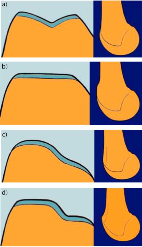
Recurrent patellar dislocation is a disabling condition, which can lead to articular cartilage injuries, osteochondral fractures, recurrent instability, pain, decreased activity and patellofemoral osteoarthritis. Trochlear dysplasia represents an important component of patellar dislocation.Imaging provides an objective basis for the morphological abnormalities and thus allows determination of the surgical strategy according to the concept of 'à la carte' surgery.The main surgical techniques of trochleoplasty are the sulcus deepening trochleoplasty, the 'Bereiter' trochleoplasty and the recession trochleoplasty.At mid-term, all techniques have shown a postoperative improvement in clinical scores, with a low rate of recurrence of dislocation and a possible return to sport. But these techniques do not halt the progression of patellofemoral arthritis. Cite this article: EFORT Open Rev 2018;3 DOI: 10.1302/2058-5241.3.170058.
Keywords: Bereiter trochleoplasty; crossing sign; deepening trochleoplasty; patellar instability; patellofemoral osteoarthritis; recession trochleoplasty; supra-trochlear spur.
Conflict of interest statement
ICMJE Conflict of interest statement: P. Neyret declares board membership of EFORT; consultancy and expert testimony for Latilini; royalties from Tornier; travel/accommodation/meetings expenses from Amplitude and Latilini, activities outside the submitted work.
Figures
References
-
- Fithian DC, Paxton EW, Stone ML, et al. Epidemiology and natural history of acute patellar dislocation. Am J Sports Med 2004;32:1114-21. - PubMed
-
- Hsiao M, Owens BD, Burks R, Sturdivant RX, Cameron KL. Incidence of acute traumatic patellar dislocation among active-duty United States military service members. Am J Sports Med 2010;38:1997-2004. - PubMed
-
- Dejour H, Walch G, Nove-Josserand L, Guier C. Factors of patellar instability: an anatomic radiographic study. Knee Surg Sports Traumatol Arthrosc 1994;2:19-26. - PubMed
-
- Dejour H, Walch G, Neyret P, Adeleine P. [Dysplasia of the femoral trochlea]. Rev Chir Orthop Repar Appar Mot 1990;76:45-54. - PubMed
LinkOut - more resources
Full Text Sources
Other Literature Sources
