

Radiological findings in ancient Egyptian canopic jars: comparing three standard clinical imaging modalities (x-rays, CT and MRI)
- PMID: 29951641
- PMCID: PMC6008346
- DOI: 10.1186/s41747-018-0048-3
Radiological findings in ancient Egyptian canopic jars: comparing three standard clinical imaging modalities (x-rays, CT and MRI)
Abstract
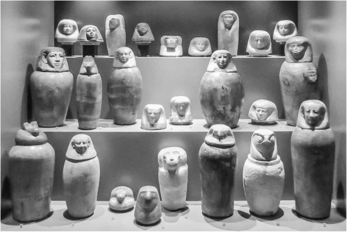
Background: The aim of our study was to evaluate the potential and the limitations of standard clinical imaging modalities for the examination of ancient Egyptian canopic jars and the mummified visceral organs (putatively) contained within them.
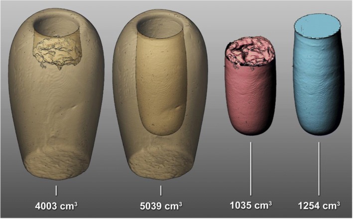
Methods: A series of four ancient Egyptian canopic jars was imaged comparing the three standard clinical imaging modalities: x-rays, computed tomography (CT) and magnetic resonance imaging (MRI). Additionally, imaging-data-based volumetric calculations were performed for quantitative assessment of the jar contents.
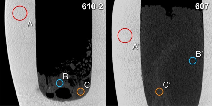
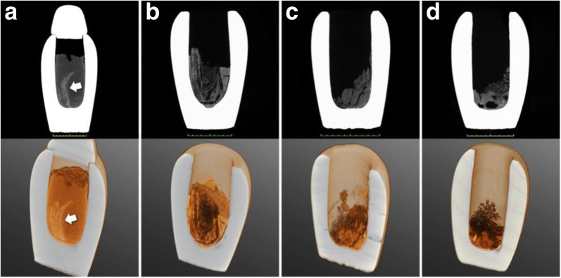
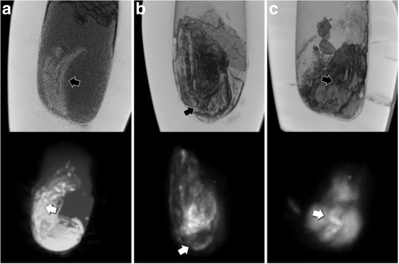
Results: The image contrast of the x-ray images was limited by the thickness and high density of the calcite mineral constituting the examined jars. CT scans showed few artefacts and revealed hyperdense structures of organ-specific morphology, surrounded by a hypodense homogeneous material. The image quality of MRI scans was limited by the low amount of water present in the desiccated jar contents. Nevertheless, areas of pronounced signal intensity coincided well with hyperdense structures previously identified on CT scans. CT-based volumetric calculations revealed holding capacities of the jars of 626-1319 cm3 and content volumes of 206-1035 cm3.
Conclusions: CT is the modality of choice for non-invasive examination of ancient Egyptian canopic jars. However, despite its limitations, x-ray imaging will often remain the only practicable method for on-site investigations. Overall, the presented radiological findings are more compatible with contained small organ fragments rather than entire mummified organs, as originally expected, with consequent implications for envisioned future sampling for chemical and genetic analysis.
Keywords: Ancient Egyptian canopic jars; Computed tomography; Magnetic resonance imaging; Paleoradiology; Radiography.
Conflict of interest statement
Not applicable.Not applicable.The authors declare that they have no competing interests.Springer Nature remains neutral with regard to jurisdictional claims in published maps and institutional affiliations.
Figures

References
-
- Koenig W, Barth JA. 14 Photographien mit Roentgen-Strahlen: aufgenommen im physikalischen Verein zu Frankfurt a.M. Verlag von Johann Ambrosius Barth, Leipzig. 1896.
-
- Lewin PK, Harwood-Nash DC (1977) Computerized axial tomography in medical archeology. Paleopathol Newsl:17–8–9 - PubMed
-
- Saab G, Chhem RK, Bohay R (2008) Paleoradiologic Techniques. Paleoradiology imaging mummies and fossils. Brothwell DR and Chhem RK. Berlin Heidelberg, Springer Verlag: 15–54
-
- Ruhli FJ, Chhem RK, Boni T. Diagnostic paleoradiology of mummified tissue: interpretation and pitfalls. Can Assoc Radiol J. 2004;55:218–227. - PubMed
LinkOut - more resources
Full Text Sources
Other Literature Sources