

Early missed abortion is associated with villous angiogenesis via the HIF-1α/VEGF signaling pathway
- PMID: 29951709
- PMCID: PMC6096576
- DOI: 10.1007/s00404-018-4802-9
Early missed abortion is associated with villous angiogenesis via the HIF-1α/VEGF signaling pathway
Abstract
Purpose: To analyze the effects of the hypoxia-inducible factor 1-alpha (HIF-1α)/vascular endothelial growth factor (VEGF) signaling pathway on villous angiogenesis in early missed abortion.
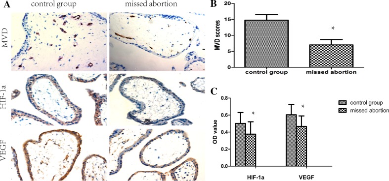
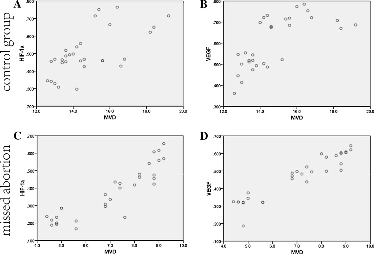
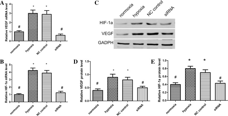
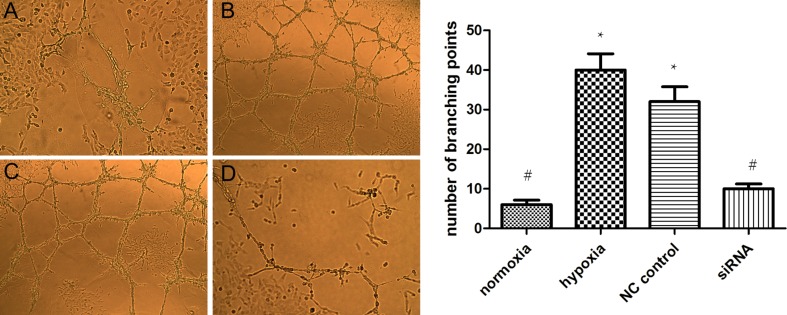
Methods: Immunohistochemical assays were performed to detect the expression of micro-vessel density (MVD), HIF-1α, and VEGF in villous tissue samples from 30 missed abortions and 30 elective abortions in early pregnancy. We further analyzed the correlation between HIF-1α/VEGF and MVD. HTR8/SVneo cells were cultured under hypoxic (1%) or normoxic (20%) conditions, tube formation was investigated, and protein and mRNA level of HIF-1α/VEGF were determined using western blot and qRT-PCR. Finally, HIF-1α was knocked down with siRNA introduced into HTR8/SVneo cell line under hypoxia, and HIF-1α/VEGF expression and HTR8/SVneo tube formation were investigated.
Results: The expression of HIF-1α, VEGF, and MVD was lower in the missed abortion than in the elective abortion group. Correlational analysis showed that the expression of HIF-1α and VEGF was positively correlated with MVD in both groups. The levels of HIF-1α/VEGF mRNA and protein in HTR8/SVneo cells were significantly enhanced under hypoxia. HIF-1α knockdown with siRNA inhibited HIF-1α/VEGF mRNA and protein levels of HTR8/SVneo cells induced by hypoxia. Tube formation of HTR8/SVneo cells was significantly enhanced in hypoxic culture and was inhibited by HIF-1α knockdown with siRNA.
Conclusions: Our results reveal a novel role for HIF-1α/VEGF in regulating villous angiogenesis in early pregnancy and suggest that it may be a novel biomarker for missed abortion.
Keywords: Angiogenesis; HIF-1α; Missed abortion; VEGF.
Conflict of interest statement
Conflict of interest
We declare that we have no conflict of interest.
Ethical approval
The study was approved by ethics committee of hospital.
Figures
References
-
- Dakouane-Giudicelli M, Brouillet S, Traboulsi W, Torre A, Vallat G, Si Nacer S, Vallee M, Feige JJ, Alfaidy N, de Mazancourt P. Inhibition of human placental endothelial cell proliferation and angiogenesis by netrin-4. Placenta. 2015;36(11):1260–1265. doi: 10.1016/j.placenta.2015.09.007. - DOI - PubMed
-
- Windsperger K, Dekan S, Pils S, Golletz C, Kunihs V, Fiala C, Kristiansen G, Knofler M, Pollheimer J. Extravillous trophoblast invasion of venous as well as lymphatic vessels is altered in idiopathic, recurrent, spontaneous abortions. Hum Reprod (Oxford, England) 2017;32(6):1208–1217. doi: 10.1093/humrep/dex058. - DOI - PubMed
Publication types
MeSH terms
Substances
LinkOut - more resources
Full Text Sources
Other Literature Sources
Medical
