Practice Guideline
. 2018 Sep;53(9):1006-1034.
doi: 10.1007/s00535-018-1484-9. Epub 2018 Jun 27.

Clinical guidelines for primary sclerosing cholangitis 2017

Affiliations
Practice Guideline

Clinical guidelines for primary sclerosing cholangitis 2017

Hiroyuki Isayama et al. J Gastroenterol. 2018 Sep.

Erratum in

  • Correction to: Clinical guidelines for primary sclerosing cholangitis 2017.
    Isayama H, Tazuma S, Kokudo N, Tanaka A, Tsuyuguchi T, Nakazawa T, Notohara K, Mizuno S, Akamatsu N, Serikawa M, Naitoh I, Hirooka Y, Wakai T, Itoi T, Ebata T, Okaniwa S, Kamisawa T, Kawashima H, Kanno A, Kubota K, Tabata M, Unno M, Takikawa H; PSC guideline committee Members: Ministry of Health, Labour and Welfare (Japan) Research Project, The Intractable Hepatobiliary Disease Study Group. Isayama H, et al. J Gastroenterol. 2022 Jun;57(6):453-454. doi: 10.1007/s00535-022-01867-7. J Gastroenterol. 2022. PMID: 35294681 Free PMC article. No abstract available.

Abstract

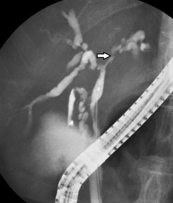
Background: Primary sclerosing cholangitis (PSC) is relatively rare disease and pathogenesis and methods of treatments were still not established. Then, we had conducted the making clinical guidelines to manage patients with PSC based on the literature review and expert opinions. These clinical guidelines were made for the medical doctors on the management of PSC, except child case of PSC.

Methods: We had employed modified Delphi method. The production committee decided guidelines, strength of recommendations and evidence level after reviewed literatures systematically, and The Expert panel evaluated those. The Scientific Committee of the Japan Biliary Association (JBA) evaluated revised guidelines, and the Public comments were collected on web site of JBA.

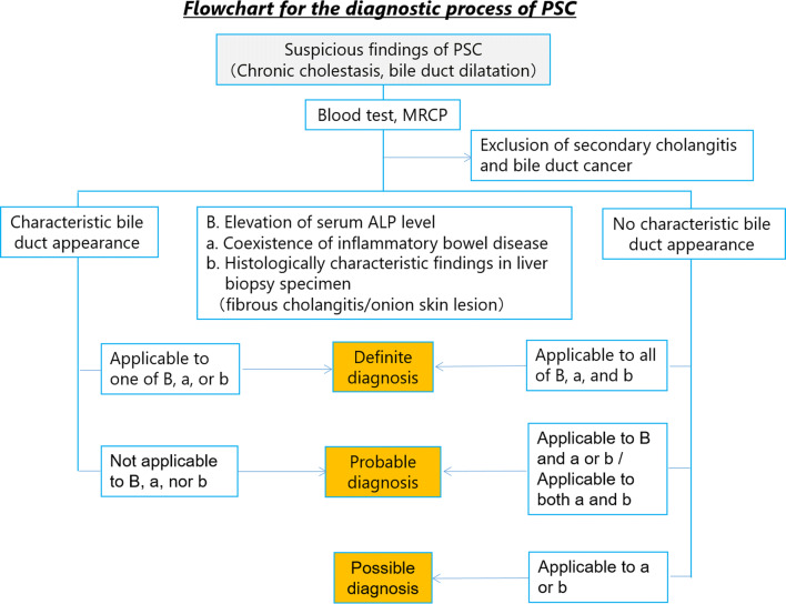
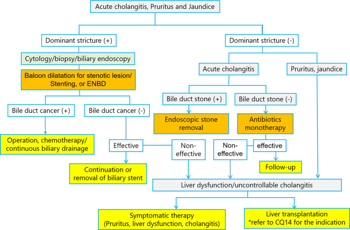
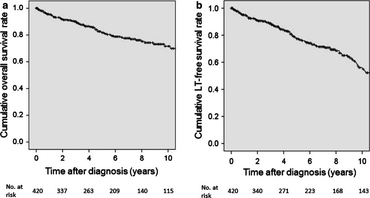
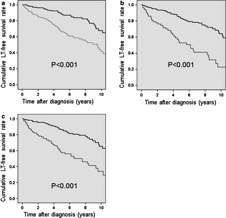
Results: We had made 16 guidelines about epidemiology/pathophysiology, diagnostics, therapy and prognosis. Also, we had made both diagnostic and therapeutic flow chart.

Conclusions: We hope that these guidelines will contribute to the improvement and development of the medical care of PSC.

Keywords: Benign biliary stricture; Cholestasis; Guidelines; Primary sclerosing cholangitis; Sclerosing cholangitis.


Conflict of interest statement

All authors declare that they have no conflict of interest.

Figures

Fig. 1
Fig. 1
Histopathological findings of the liver indicating PSC (Modified citation from Ref. [18]). Characteristics include ring-like fibrosis surrounding the bile duct and inflammatory cell infiltration. Concentric, nested fibrosis that resembles and onion and is therefore known as “onion-skin fibrosis”—is presented
Fig. 2
Fig. 2
Distribution of age at diagnosis for PSC patients in Japan (Modified citation from Ref. [30])
Fig. 3
Fig. 3
Distribution of age at diagnosis of PSC in Northern California (Modified citation from Ref. [27])
Fig. 4
Fig. 4
Flowchart of diagnostic strategies of PSC
Fig. 5
Fig. 5
Ultrasound Image of PSC. a Common bile duct wall thickening is observed. (right intercostal scanning). b Intrahepatic bile duct at the umbilical region are dilated and have thickened irregularly. (wide probe scanning)
Fig. 6
Fig. 6
MRCP Image of PSC. a Alternations between strictures and slightly dilated segments produce a beaded pattern. (white arrow). b This image shows non-stricture of the common bile duct in the part of the duodenal side from the cystic duct, it also demonstrates gallbladder enlargement. This enlargement is non-specific but refers to findings of PSC
Fig. 7
Fig. 7
Colonoscopy image of the inflammatory bowel disease accompanied with PSC. a This picture shows multiple ulcerative stenosis in the area from the ileocecal to the ascending colon, but no rectal lesion has been found. (at the time of this diagnosis). b Multiple erosion and vascular permeability reduction have appeared in the same area (7 months after the diagnosis)
Fig. 8
Fig. 8
Characteristic bile duct image of PSC. a Band-like stricture findings with ERC. Multifocal, short, annular strictures can be observed in the intrahepatic ducts (white arrows). b Beaded appearance findings with ERC. Alternations between strictures and slightly dilated segments produce a beaded pattern (white arrows). c Pruned tree appearance findings with ERC: Early stage. Overall intrahepatic branch ducts are narrow but can be detected with ERC. d Pruned tree appearance findings with ERC: advanced stage. Branch ducts seem to have disappeared as if they were pruned trees. e Diverticulum-like outpouching findings with ERC. Extrahepatic bile ducts have outpouching diverticular-like appearance (white arrow)
Fig. 9
Fig. 9
Comparison Between PSC and IgG4-SC with the bile duct image. (Modified citation from Ref. [89]). a Primary sclerosing cholangitis. (1) Band-like stricture, (2) beaded appearance, (3) pruned tree appearance, (4) diverticulum-like outpouching, (5) shaggy appearance. b IgG4-related SC. (6) Dilation after confluent stricture, (7) stricture of lower common bile duct, (8) stricture of hepatic hilar
Fig. 10
Fig. 10
Intraductal ultrasound (IDUS) findings. a IDUS of PSC: this image shows an irregularity of the bile duct epithelium and diverticulum-like outpouching (white arrow). b IDUS of IgG4-related SC: bile duct epithelium is smooth and preserves three-layer structure. Regular hypoechoic thickening in the inner part can be seen
Fig. 11
Fig. 11
Peroral cholangioscopy (POCS) findings of PSC. a Ulcerative scars. b Multiple diverticulum-like findings
Fig. 12
Fig. 12
Small duct disappearance and periportal ductular proliferation
Fig. 13
Fig. 13
ERCP image of dominant stricture. A bile duct stricture within 2 cm of the branch between the right and left hepatic ducts can be seen
Fig. 14
Fig. 14
Flowchart for the management of symptomatic PSC
Fig. 15
Fig. 15
Prognosis of PSC patients in Japan (Modified citation from Ref. [30]). a Overall survival. b Overall survival without liver transplantation
Fig. 16
Fig. 16
Prognosis of PSC patients in Japan: Stratification based on prognostic factors. (Modified citation from a 2015 National Survey on PSC: Ref. [30]). a Age at diagnosis: bold line, < 44 years; thin line, ≥ 44 years. b Serum albumin level at diagnosis: bold line, ≥ 3.5 g/dl; thin line, < 3.5 g/dl. c Total bilirubin level at diagnosis: bold line, < 1.5 mg/dl; thin line, ≥ 1.5 mg/dl

References

    1. Lindor KD, LaRusso NF. Primary sclerosing cholangitis. In: Schiff ER, editor. Disease of the liver. 9. Philadelphia: Lippincott Williams & Wilkins; 2003. pp. 673–684.
    1. Tabibian JH, O’Hara SP, Lindor KD. Primary sclerosing cholangitis and the microbiota: current knowledge and perspectives on etiopathogenesis and emerging therapies. Scand J Gastroenterol. 2014;49:901–908. doi: 10.3109/00365521.2014.913189. - DOI - PMC - PubMed
    1. Aron JH, Bowlus CL. The immunobiology of primary sclerosing cholangitis. Semin Immunopathol. 2009;31:383–397. doi: 10.1007/s00281-009-0154-7. - DOI - PMC - PubMed
    1. O’Mahony CA, Vierling JM. Etiopathogenesis of primary sclerosing cholangitis. Semin Liver Dis. 2006;26:3–21. doi: 10.1055/s-2006-933559. - DOI - PubMed
    1. Eaton JE, Talwalkar JA, Lazaridis KN, et al. Pathogenesis of primary sclerosing cholangitis and advances in diagnosis and management. Gastroenterology. 2013;145:521–536. doi: 10.1053/j.gastro.2013.06.052. - DOI - PMC - PubMed

Publication types

MeSH terms

LinkOut - more resources