Review
. 2019 Jun;176(12):1818-1828.
doi: 10.1111/bph.14427. Epub 2018 Jul 20.

Role of immune cells in hypertension

Affiliations
Review

Role of immune cells in hypertension

Antoine Caillon et al. Br J Pharmacol. 2019 Jun.

Abstract

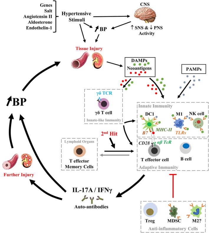
Inflammatory processes have been shown to play an important role in the mechanisms involved in the pathogenesis of hypertension. Innate and adaptive immune responses participate in BP elevation and end-organ damage. Here, we discuss recent studies focusing on novel inflammatory and immune mechanisms that play roles in BP elevation. Different subpopulations of cells involved in innate and adaptive immune responses, such as dendritic cells, monocytes/macrophages and NK cells, on the one hand, and B and T lymphocytes, on the other, contribute to the vascular and kidney injury in hypertension. Unconventional innate-like T cells such as γδ T cells also participate in hypertensive mechanisms by priming both innate and adaptive immune cells, contributing to trigger vascular inflammation and BP elevation. These cells exert their effects in part via production of various cytokines including pro-inflammatory IFN-γ and IL-17 and anti-inflammatory IL-10. The present review summarizes some of these immune mechanisms that participate in the pathophysiology of hypertension. LINKED ARTICLES: This article is part of a themed section on Immune Targets in Hypertension. To view the other articles in this section visit http://onlinelibrary.wiley.com/doi/10.1111/bph.v176.12/issuetoc.


Conflict of interest statement

The authors declare no conflicts of interest.

Figures

Figure 1
Figure 1
Role of immune cells in inflammation in hypertension. Environmental factors such as salt, in the presence of genetic susceptibility, may lead to small rises of BP, in part, by triggering the activation of the sympathetic nervous system (SNS) and inhibition of the parasympathetic nervous system (PNS) alone or together with other hypertensive stimuli such as Ang II, aldosterone and endothelin‐1. Over time, high BP and/or the pro‐hypertensive stimuli induce tissue injury, which together with oxidative stress caused by vasoactive peptides such as Ang II or endothelin‐1 create favourable conditions for the development of DAMPs and neoantigens, such as isoketal protein adducts. DAMPs activate innate immunity via TLRs on type 1 macrophages (M1), type 1 dendritic cells (DC1) and NK cells, while neoantigens enhance DC immunogenicity and promote DC release of IL‐6, IL‐1β and IL‐23, causing proliferation of T cells and production of IL‐17A, IFN‐γ and TNF‐α. DAMPs and neoantigens could also activate unconventional lymphocytes such as innate‐like γδ T cells, which will activate T lymphocytes. Opportunistic diseases such as periodontitis could also exacerbate the activation of the innate immune system through binding of PAMPs to pattern recognition receptors such as TLRs. Innate immune cells and γδ T cells contribute to inflammation, both directly or via the activation of adaptive immunity, inducing pro‐inflammatory cytokines, such as IL‐17 and IFN‐γ, and the production of autoantibodies, leading to vascular and kidney injury, which closes the pro‐hypertensive circle that is a feed‐forward process resulting in progressive BP elevation. During this process, TEM cells are produced and stored in lymphoid organs including the bone marrow. Upon a second hit such as high‐salt intake, TEM cells could be reactivated and contribute to the vicious cycle to maintain high BP or further increase its level. Throughout this process, anti‐inflammatory cells such as Treg, MDSCs and type 2 macrophages (M2) could provide homeostatic fine‐tuning of the inflammatory process in blood vessels and the kidney. However, this anti‐inflammatory mechanism could be rendered unable to counteract the development of hypertension because of factors such as Ang II that can cause a decrease in the number and function of Treg through activation of the complement system. B7, co‐stimulatory molecule on antigen presenting cell surface (CD80 or CD86).

References

    1. Alexander SP, Christopoulos A, Davenport AP, Kelly E, Marrion NV, Peters JA et al (2017a). The concise guide to pharmacology 2017/18: G protein‐coupled receptors. Br J Pharmacol 174 (Suppl 1): S17–S129. - PMC - PubMed
    1. Alexander SP, Cidlowski JA, Kelly E, Marrion NV, Peters JA, Faccenda E et al (2017b). The concise guide to pharmacology 2017/18: Nuclear hormone receptors. Br J Pharmacol 174 (Suppl 1): S208–S224. - PMC - PubMed
    1. Alexander SP, Fabbro D, Kelly E, Marrion NV, Peters JA, Faccenda E et al (2017c). The concise guide to pharmacology 2017/18: Catalytic receptors. Br J Pharmacol 174 (Suppl 1): S225–S271. - PMC - PubMed
    1. Alexander SP, Fabbro D, Kelly E, Marrion NV, Peters JA, Faccenda E et al (2017d). The concise guide to pharmacology 2017/18: Enzymes. Br J Pharmacol 174 (Suppl 1): S272–S359. - PMC - PubMed
    1. Alexander SP, Peters JA, Kelly E, Marrion NV, Faccenda E, Harding SD et al (2017e). The concise guide to pharmacology 2017/18: Ligand‐gated ion channels. Br J Pharmacol 174 (Suppl 1): S130–S159. - PMC - PubMed

Publication types