. 2018 Aug 17;13(8):2347-2358.
doi: 10.1021/acschembio.8b00568. Epub 2018 Jul 11.

Parathyroid Hormone Senses Extracellular Calcium To Modulate Endocrine Signaling upon Binding to the Family B GPCR Parathyroid Hormone 1 Receptor

Affiliations

Parathyroid Hormone Senses Extracellular Calcium To Modulate Endocrine Signaling upon Binding to the Family B GPCR Parathyroid Hormone 1 Receptor

Kelly J Culhane et al. ACS Chem Biol. .

Abstract

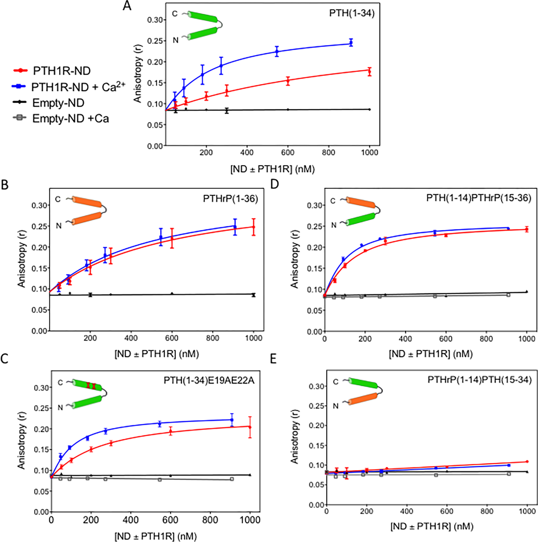
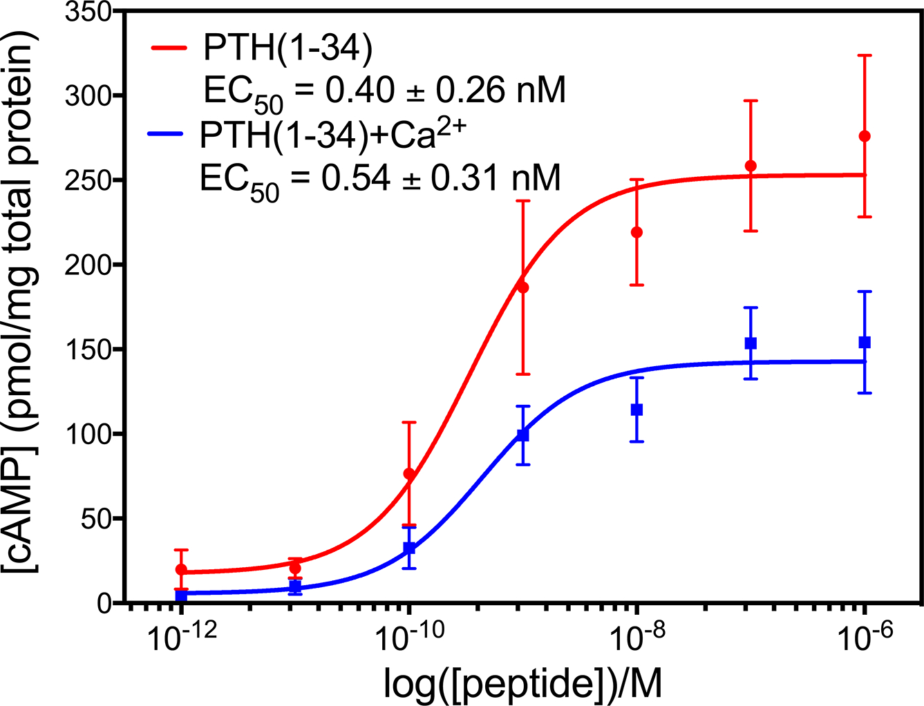
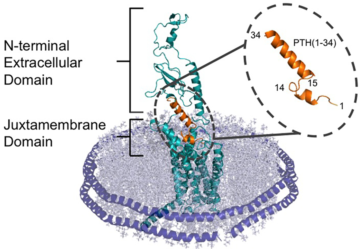
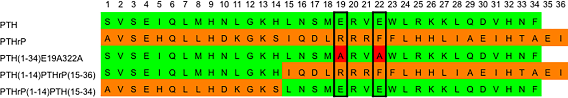
Parathyroid hormone (PTH) binds to a family B G protein coupled receptor, parathyroid hormone 1 receptor (PTH1R). One of its functions is to regulate Ca2+ homeostasis in bone remodeling, during which Ca2+ can reach up to 40 mM. A truncated version of PTH, PTH(1-34), can fully activate PTH1R and has been used for osteoporosis treatments. Here, we used fluorescence anisotropy to examine the binding of PTH(1-34) to PTH1R purified in nanodiscs (PTH1R-ND) and found that the affinity increases 5-fold in the presence of 15 mM Ca2+. However, PTHrP(1-36), another truncated endogenous agonist for PTH1R, does not show this Ca2+ effect. Mutations of Glu19 and Glu22 in PTH(1-34) that are not conserved in PTHrP(1-36) largely abolished the Ca2+ effect. The results support that PTH(1-34) not only activates PTH1R but also uniquely senses Ca2+. This dual function of a peptide hormone is a novel observation that couples changes in extracellular environment with endocrine signaling. Understanding this can potentially reveal the complex role of PTH signaling in bone remodeling and improve the PTH(1-34) treatment for osteoporosis.


Figures

Figure 1:
Figure 1:. Binding of peptide ligands to PTH1R incorporated in nanodiscs (PTH1R-ND)
Titrations of (A) PTH(1–34); (B) PTHrP(1–36); (C) PTH(1–34)E19AE22A; (D) PTH(1–14)PTHrP(15–36) and (E) PTHrP(1–14)PTH(15–34) with PTH1R in nanodiscs (PTH1R-ND) with (blue) and without (red) addition of 15 mM Ca2+ and nanodiscs containing no PTH1R (empty-ND) with (grey) and without (black) addition of 15 mM Ca2+. The peptide concentration was kept constant at 50 and 55 nM with and without addition of 15 mM Ca2+, respectively. (Buffer: 50 mM Tris-HCl pH 7.4, 150 mM NaCl, 3 mM MgCl2, and 100 μM EDTA). Each data point in the titration curves is an average of more than three experiments and each experiment was performed using independent preparations of purified PTH1R-ND.
Figure 2:
Figure 2:. Flow cytometry analysis of PTH(1–34)-FAM binding to HEK293S cells expressing PTH1R.
Titrations of HEK293S cells expressing PTH1R with PTH(1–34)-FAM in the presence of 0 mM Ca2+ (red) and 15 mM Ca2+ (blue) and HEK293S cells with no induced PTH1R expression in the presence of 0 mM Ca2+ (grey) and 15 mM Ca2+ (black). Data shown are representative curves from of one of five replicates (SI Figure S3).
Figure 3:
Figure 3:. Binding of PTH(1–34) to the extracellular domain (ECD) of PTH1R.
(A) Construction of MBP-PTH1R-ECD. (B) SDS-PAGE gel of MBP-PTH1R-ECD shows Lane 1, markers; lane 2, purified MBP-PTH1R-ECD (62 kD) (C) Binding of PTH(1–34) to MBP-PTH1R-ECD measured by fluorescence polarization. (Buffer: 50 mM Tris-HCl, pH 7.5, 150 mM NaCl, 1 mM EDTA). Each curve shows the average of three fluorescence polarization experiments.
Figure 4:
Figure 4:. Specific Ca2+ effect on PTH(1–34) binding to PTH1R-ND.
Titration of PTH(1–34)-FAM at 50 nM with PTH1R-ND in Mg2+-depleted buffer (50 mM Tris-HCl pH 7.4, 150 mM NaCl, and 100 μM EDTA) at various concentration of (A) Mg2+ and (B) Ca2+. Unless specified, each curve shows the average of at least three replicates. The 10 mM Mg2+, 40 mM Mg2+ and 10 mM Ca2+ titrations show a single experiment.
Figure 5:
Figure 5:. PTH(1–34) activates PTH1R to produce cAMP in the presence and absence of 15 mM Ca2+.
HEK293S cells expressing PTH1R were stimulated with indicated concentrations of PTH(1–34) with 0 mM Ca2+ (red) and 15 mM Ca2+ (blue) for 30 minutes before harvesting to measure the concentration of cAMP. The addition of 15 mM Ca2+ does not change the potency of PTH(1–34) but does decrease the efficacy of PTH(1–34). Titration curves show the average of 3 experiments.
Scheme 1:
Scheme 1:. PTH(1–34) bound to PTH1R shown in a nanodisc.
The C terminus of PTH(1–34) (orange) interacts with the extracellular domain of the receptor, while the N terminus of the peptide hormone interacts with the juxtamembrane domain. The receptor is surround by a lipid bilayer (grey), which is held together with the alpha helical membrane scaffold protein MSP1E3D1 (purple).
Scheme 2:
Scheme 2:. Sequence alignment of the PTH and PTHrP ligands under study.
PTH residues (green), PTHrP residues (orange), and E19A and E22A mutations (red).
Scheme 3:
Scheme 3:. Extracellular domain of PTH1R has a high density of negatively charged residues.
Snake plot of PTH1R shows negatively charged residues in red, which are clustered towards the extracellular side of the receptor. Two regions of PTH1R not conserved in family B GPCRs are boxed: the 50-residue flexible loop contains 8 negatively charged residues and the 16-residue extracellular loop 1 contains 6 negatively charged residues.

References

    1. Stevens RC, Cherezov V, Katritch V, Abagyan R, Kuhn P, Rosen H, and Wüthrich K (2013) The GPCR Network: a large-scale collaboration to determine human GPCR structure and function, Nat. Rev. Drug Discovery 12, 25–34. - PMC - PubMed
    1. Culhane KJ, Liu Y, Cai Y, and Yan EC (2015) Transmembrane signal transduction by peptide hormones via family BG protein-coupled receptors, Front. Pharmacol. 6. - PMC - PubMed
    1. Hollenstein K, de Graaf C, Bortolato A, Wang M-W, Marshall FH, and Stevens RC (2014) Insights into the structure of class B GPCRs, Trends in Pharmacol. Sci. 35, 12–22. - PMC - PubMed
    1. Pal KM, Karsten: Xu H Eric. (2012) Structure and mechanism for recognition of peptide hormones by Class B G protein coupled receptors, Acta Pharmacol. Sin. 33, 300–311. - PMC - PubMed
    1. Liang YL, Khoshouei M, Radjainia M, Zhang Y, Glukhova A, Tarrasch J, Thal DM, Furness SGB, Christopoulos G, Coudrat T, Danev R, Baumeister W, Miller LJ, Christopoulos A, Kobilka BK, Wootten D, Skiniotis G, and Sexton PM (2017) Phase-plate cryo-EM structure of a class B GPCR-G-protein complex, Nature. - PMC - PubMed

Publication types