

CT texture analysis of histologically proven benign and malignant lung lesions
- PMID: 29952966
- PMCID: PMC6039644
- DOI: 10.1097/MD.0000000000011172
CT texture analysis of histologically proven benign and malignant lung lesions
Abstract
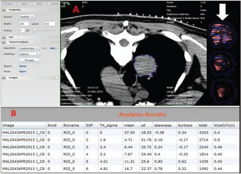
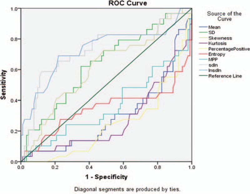
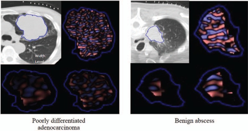
The purpose of our study was to determine accuracy of CT texture analysis (CTTA) for differentiating benign from malignant pulmonary nodules, and well-differentiated from poorly differentiated lung cancers, with histology as the standard of reference.In this IRB-approved study, 175 adult patients (average age 66 ± 12 years; age range 27-89 years, male 82: female 93) who underwent a noncontrast chest CT examination prior to CT-guided biopsy of pulmonary nodules were included. There were 57 benign (24 tumors or tumor-like lesions; 33 inflammatory conditions) and 120 malignant (29 well-differentiated adenocarcinomas, 48 poorly differentiated adenocarcinomas, and 43 squamous cell carcinomas) diagnoses on pathology. CTTA was performed on the prebiopsy noncontrast CT images using a commercially available software (TexRAD limited, UK). The CTCA features analyzed included mean HU values, percent positive pixels (PPP), mean value of positive pixels (MPP), standard deviation (SD), normalized SD, skewness, kurtosis, and entropy.The ROC analyses showed that normalized SD [AUC: 0.63, (CI: 0.55-72), P = .003] had moderate accuracy for differentiating between benign and malignant lesions. For differentiating among well-differentiated and poorly differentiated tumors, the ROC analysis showed that except skewness all other parameters were statistically significant The AUC values of other CTTA parameters were: mean (AUC: 0.73-0.76, P = .001- < .0001).CT texture analyses can reliably predict well- and poorly differentiated lung malignancies. However, inflammatory lung lesions with tissue heterogeneity negatively affect the performance of CTTA when it comes to differentiation between benign and malignant pulmonary nodules.
Conflict of interest statement
The authors have no conflicts of interest to disclose.
Figures

References
-
- Ozeki N1, Iwano S2, Taniguchi T1, et al. Therapeutic surgery without a definitive diagnosis can be an option in selected patients with suspected lung cancer. Interact Cardiovasc Thorac Surg 2014;19:830–7. - PubMed
-
- Merritt RE, Shrager JB. Indications for surgery in patients with localized pulmonary infection. Thorac Surg Clin 2012;22:325–32. - PubMed
-
- Scott WJ. Surgical treatment of other bronchial tumors. Chest Surg Clin N Am 2033;13:111–2. - PubMed
MeSH terms
LinkOut - more resources
Full Text Sources
Other Literature Sources
Medical
