Observational Study
. 2018 Jun;97(26):e11195.
doi: 10.1097/MD.0000000000011195.

Discrimination of pyogenic spondylitis from brucellar spondylitis on MRI

Affiliations
Observational Study

Discrimination of pyogenic spondylitis from brucellar spondylitis on MRI

Tao Li et al. Medicine (Baltimore). 2018 Jun.

Abstract

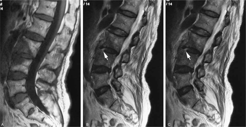
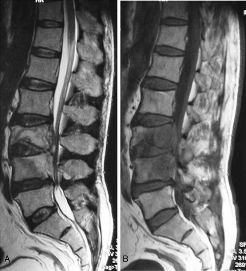
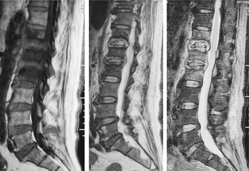
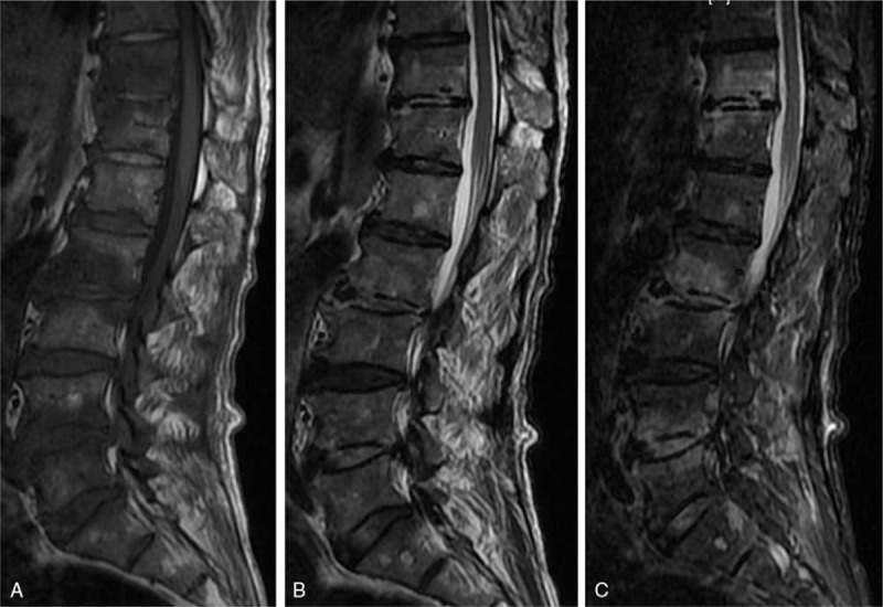
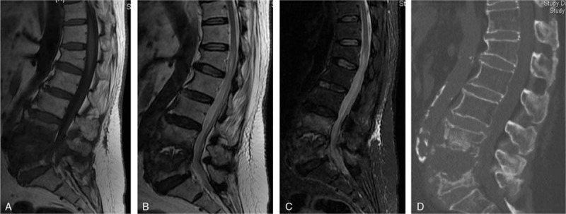
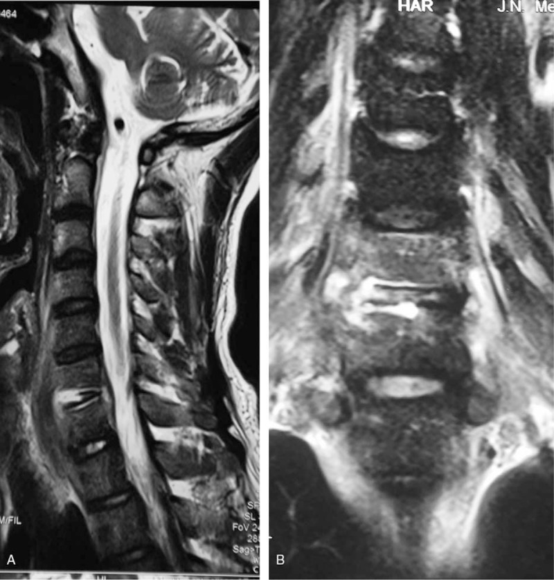
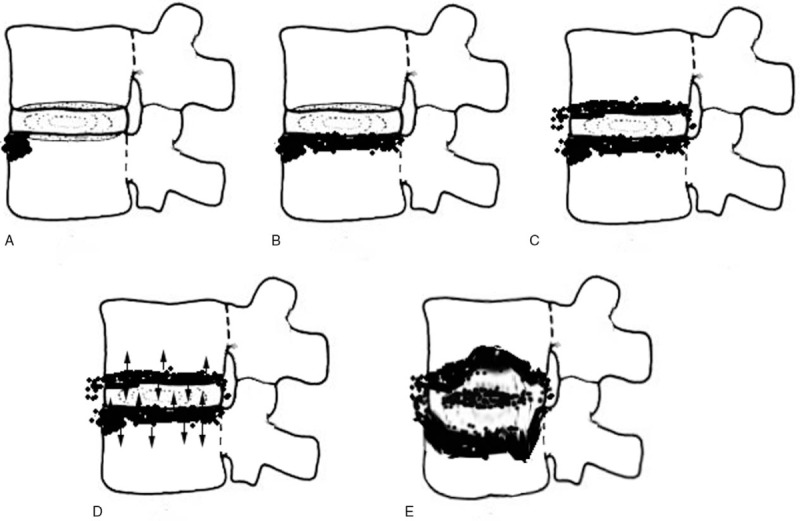
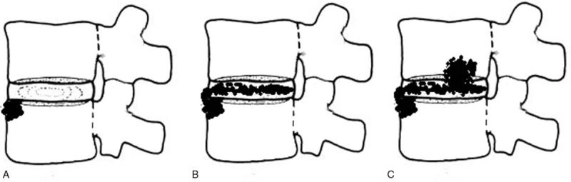
Retrospective Cross-Sectional Study.The purpose of this study was to investigate the accuracy of magnetic resonance imaging (MRI) for distinguishing between pyogenic spondylitis and brucellar spondylitis.Although pyogenic spondylodiscitis (PS) and brucellar spondylitis (BS) are common causes of spinal infections, the variety of their clinical manifestations complicates differential diagnosis. MRI may be helpful in differential diagnosis and treatment.MRI images of 64 patients who underwent MRI of the spine and with confirmed spondylitis were retrospectively reviewed. After referring to the related medical literature, we compared 32 patients with pyogenic spondylitis and 32 patients with brucellar spondylitis regarding MRI findings. Statistical analysis was performed with the chi-square test. Statistical significance was defined as P < .05.The significant differences between PS and BS on MRI findings are listed as follows (P < .05): diffuse, partial and fan-shaped hyperintense signals on middle sagittal fat-suppressed weighted images (PS: 51, 11, 3/65 vs BS:35, 18, 19/72); focal endplate destruction (PS: 9/43 vs BS:27/35); extensive end plate destruction (PS: 29/43 vs BS:8/35); ballooning change of the intravertebral space (PS: 7/32 vs BS:0/32); an inflammatory reaction line from the end plate (PS: 30/65 vs BS: 1/72); a disc invasion sign (PS: 1/28 vs BS:12/33); an inflammatory reaction line in the disc (PS: 5/28 vs BS:25/33); and 8) severe intravertebral space destruction (PS: 17/28 vs BS:12/33);MRI imaging provides useful information for the differentiation between pyogenic spondylitis and brucellar spondylitis.


Conflict of interest statement

The authors have no conflicts of interest to disclose.

Figures

Figure 1
Figure 1
A 69-year-old woman with a brucella infection for 2.5 months without any previous antibiotic treatment. (A) T1-weighted image shows hypointensity of the L3-L4 vertebral bodies, showing evident front fan-shape shade. (B and C) T2-weighted and FS MRIs show infection starting from the anterior of the endplate in both L3 and L4 and then involving the disc from L4 vertebra, which we called the Disc Invasion Sign (arrow), and invading the vertebral body (in fan shape).
Figure 2
Figure 2
A 55-year-old man with a brucella infection for 3 months without any previous antibiotic treatment. (A) T1-weighted image shows a hypointense signal from L3-L4. (B) T2-weighted image shows a hyperintense signal from L3 to L4; both (A) and (B) present posterior fan shape spreading to the vertebral body.
Figure 3
Figure 3
A 67-year-old man with a pyogenic infection (S aureus) for 2 months without any previous antibiotic treatment. A sagittal T1-weighted image (A) shows hypointensity on T12, L1 and L2. T2-weighted image (B) and FS MRIs (C) show the “inflammatory reaction line from the end plate” and the involved discs of T12, L1 and L2, which show hyperintense signals. The endplates were destroyed extensively. This infected disc and endplate comprise a typical “eye sign.” The infected disc can be thought of as an eyeball and the infected endplate can be regarded as an eyelid.
Figure 4
Figure 4
A 61-year-old man with a brucella infection for 2 months without any previous antibiotic treatment. A multiple-level involvement case. (A) T1-weighted image shows a hypointense signal from the T11-L5 vertebrae. (B) T2-weighted image shows a heterogeneous signal from the T11-L5 vertebrae. (C) FS MRI shows a hyperintense signal from the involved vertebrae. An inflammatory reaction line in the disc was found from the T11/12 and L2/3 discs. Focal endplate destruction was demonstrated from the inferior endplate of the T11 vertebra.
Figure 5
Figure 5
A 77-year-old woman with a pyogenic infection (E coli) for 1 month. (A) T1-weighted MRI shows a hypointense signal from the interspace of L4/5 and the L4 and L5 vertebrae. (B) isointensity signal are shown from L4 and L5 vertebrae. A heterogeneous signal can be observed from the L4/5 interspace, which indicates abscess formation. (C) FS-MRI shows a hyperintense signal from the L4 and L5 vertebrae. (D) The L5 vertebra has been destroyed severely, and the interspace has expanded, which we can define as “ballooning change of the intravertebral space.”
Figure 6
Figure 6
A 37-year-old man with a pyogenic infection (S aureus) for 1.5 months without any previous antibiotic treatment. The sagittal T2-weighted image (A) and coronal T2-weighted image (B) show the “inflammatory reaction line from the end plate” of C6 and C7 and the disc of C6/7 involving the infected endplates.
Figure 7
Figure 7
A simulated image of the infection course in PS. (A) The infection usually started from the front edge of the endplate. (B) The endplate was infected in line. (C) The next endplate was infected. (D) Both involved endplates further infected the vertebrae and disc, respectively (arrow). (E) With further destruction, the intravertebral space expands toward a “ballooning change.”
Figure 8
Figure 8
A simulated image of the infection course between the disc and vertebrae in BS. (A) The infection usually started from the front edge of the endplate. The shaded area shows the endplate edema. (B) The infection was involved into the disc (shadow on disc). (C) The infected disc further involves the endplate of next vertebra and causes focal destruction from the endplate.

References

    1. Baleriaux DL, Neugroschl C. Spinal and spinal cord infection. Eur Radiol 2004;14(suppl 3):E72–83. - PubMed
    1. Tali ET. Spinal infections. Eur J Radiol 2004;50:120–33. - PubMed
    1. Mukherji SK. Spinal infections. Neuroimaging Clin N Am 2015;25:xiii. - PubMed
    1. Gold M. Magnetic resonance imaging of spinal emergencies. Top Magn Reson Imaging 2015;24:325–30. - PubMed
    1. Tali ET, Koc AM, Oner AY. Spinal brucellosis. Neuroimag Clin N Am 2015;25:233–45. - PubMed

Publication types