. 2018 Aug;8(8):e01028.
doi: 10.1002/brb3.1028. Epub 2018 Jun 28.

Gunn rats with glial activation in the hippocampus show prolonged immobility time in the forced swimming test and tail suspension test

Affiliations

Gunn rats with glial activation in the hippocampus show prolonged immobility time in the forced swimming test and tail suspension test

Ryosuke Arauchi et al. Brain Behav. 2018 Aug.

Abstract

Introduction: Recent studies imply that glial activation plays a role in the pathogenesis of psychiatric disorders, such as schizophrenia and major depression. We previously demonstrated that Gunn rats with hyperbilirubinemia show congenital gliosis and schizophrenia-like behavior.

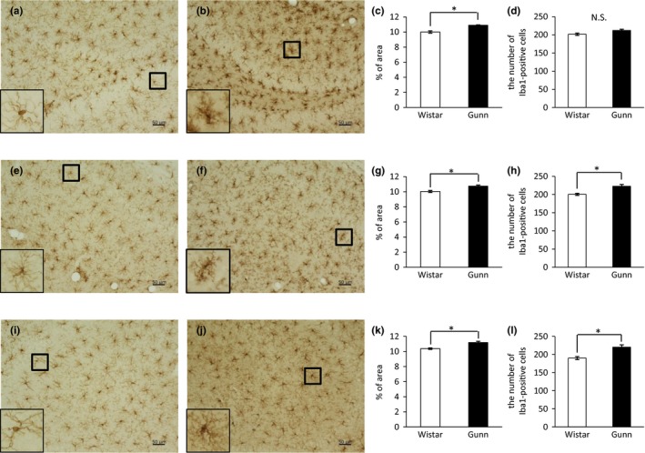
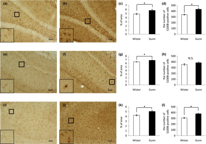
Methods: As it has been suggested that major depression involves glial activation associated with neuroinflammation, we examined whether Gunn rats show depression-like behavior using the forced swimming test (FST) and the tail suspension test (TST). In addition, we quantitatively evaluated both microgliosis and astrogliosis in the hippocampus of Gunn rats using immunohistochemistry analysis of the microglial marker ionized calcium-binding adaptor molecule (Iba) 1 and the astrocytic marker S100B.

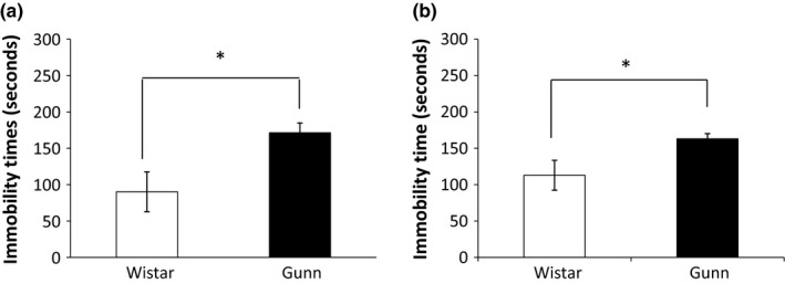
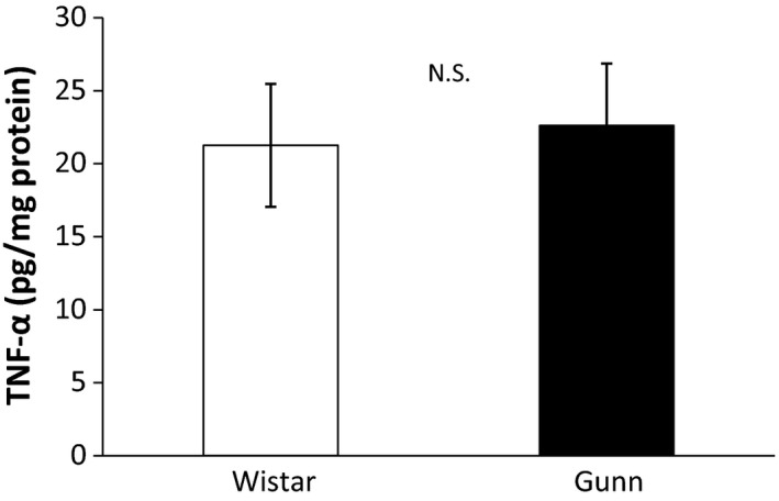
Results: Both the FST and TST showed that immobility time of Gunn rats was significantly longer than that of normal control Wistar rats, indicating that Gunn rats are somewhat helpless, a sign of depression-like behavior. In the quantification of immunohistochemical analysis, Iba1immunoreactivity in the dentate gyrus (DG), cornu ammonis (CA) 1, and CA3 and the number of Iba1-positive cells in the CA1 and CA3 were significantly increased in Gunn rats compared to Wistar rats. S100B immunoreactivity in the DG, CA1, and CA3 and the number of S100B-positive cells in the DG and CA3 were significantly increased in Gunn rats compared to Wistar rats.

Conclusion: Our findings suggest that both microglia and astrocyte are activated in Gunn rats and their learned helplessness could be related to glial activation.

Keywords: Gunn rat; astrocytes; forced swimming test; hippocampus; microglia; tail suspension test.


Figures

Figure 1
Figure 1
Immobility time of Wistar and Gunn rats in the FST and TST. Immobility time of Gunn rats is significantly longer than that of Wistar rats in both the FST (a) and the TST (b). Each value is the mean ± SEM (= 6). *< 0.05. FST, forced swimming test; TST, tail suspension test
Figure 2
Figure 2
Immunoreactivity for Iba1 in the hippocampus. Representative images of DAB staining in the DG of Wistar (a) and Gunn rats (b), CA1 of Wistar (e) and Gunn rats (f), and CA3 of Wistar (i) and Gunn rats (j). The magnified pictures of Iba1‐positive cells are shown in the lower‐left insets. The scale bars indicate 50 μm. Quantification data for Iba1 immunoreactivity in the DG (c), CA1 (g), and CA3 (k). The number of Iba1‐positive cells in the DG (d), CA1 (h), and CA3 (l). Each value is the mean ± SEM (= 6). *< 0.05. N.S., not significant
Figure 3
Figure 3
Immunoreactivity for S100B in the hippocampus. Representative images of DAB staining in the DG of Wistar (a) and Gunn rats (b), CA1 of Wistar (e) and Gunn rats (f), and CA3 of Wistar (i) and Gunn rats (j). The magnified pictures of S100B‐positive cells are shown in the lower‐left insets. The scale bars indicate 50 μm. Quantification data for S100B immunoreactivity in the DG (c), CA1 (g), and CA3 (k). The number of S100B‐positive cells in the DG (d), CA1 (h), and CA3 (l). Each value is the mean ± SEM (= 6). *< 0.05. N.S., not significant
Figure 4
Figure 4
Quantification of TNF‐α in the hippocampus. The amount of TNF‐α in the hippocampus was measured using ELISA. There was no significant difference between Gunn rats and Wistar rats. Each value is the mean ± SEM (= 8). N.S., not significant

References

    1. Bayer, T. A. , Busleia, R. , Havasb, L. , & Falkaia, P. (1999). Evidence for activation of microglia in patients with psychiatric illnesses. Neuroscience Letters, 271, 126–128. 10.1016/S0304-3940(99)00545-5 - DOI - PubMed
    1. Bernstein, H. G. , Steiner, J. , Guest, P. C. , Dobrowolny, H. , & Bogerts, B. (2015). Glial cells as key players in schizophrenia pathology: Recent insights and concepts of therapy. Schizophrenia Research, 161(1), 4–18. 10.1016/j.schres.2014.03.035 - DOI - PubMed
    1. Biesmans, S. , Meert, T. F. , Bouwknecht, J. A. , Acton, P. D. , Davoodi, N. , De Haes, P. , … Nuydens, R. (2013). Systemic immune activation leads to neuroinflammation and sickness behavior in mice. Mediators of Inflammation, 2013, 271359 10.1155/2013/271359 - DOI - PMC - PubMed
    1. Burke, N. N. , Geoghegan, E. , Kerr, D. M. , Moriarty, O. , Finn, D. P. , & Roche, M. (2013). Altered neuropathic pain behaviour in a rat model of depression is associated with changes in inflammatory gene expression in the amygdala. Genes, Brain and Behavior, 12(7), 705–713. 10.1111/gbb.12080 - DOI - PubMed
    1. Catts, V. S. , Wong, J. , Fillman, S. G. , Fung, S. J. , & Shannon Weickert, C. (2014). Increased expression of astrocyte markers in schizophrenia: Association with neuroinflammation. Australian & New Zealand Journal of Psychiatry, 48(8), 722–734. 10.1177/0004867414531078 - DOI - PubMed

Publication types