. 2018 Oct:50:48-57.
doi: 10.1016/j.cellsig.2018.06.012. Epub 2018 Jun 25.

RhoA regulates Drp1 mediated mitochondrial fission through ROCK to protect cardiomyocytes

Affiliations

RhoA regulates Drp1 mediated mitochondrial fission through ROCK to protect cardiomyocytes

Cameron S Brand et al. Cell Signal. 2018 Oct.

Abstract

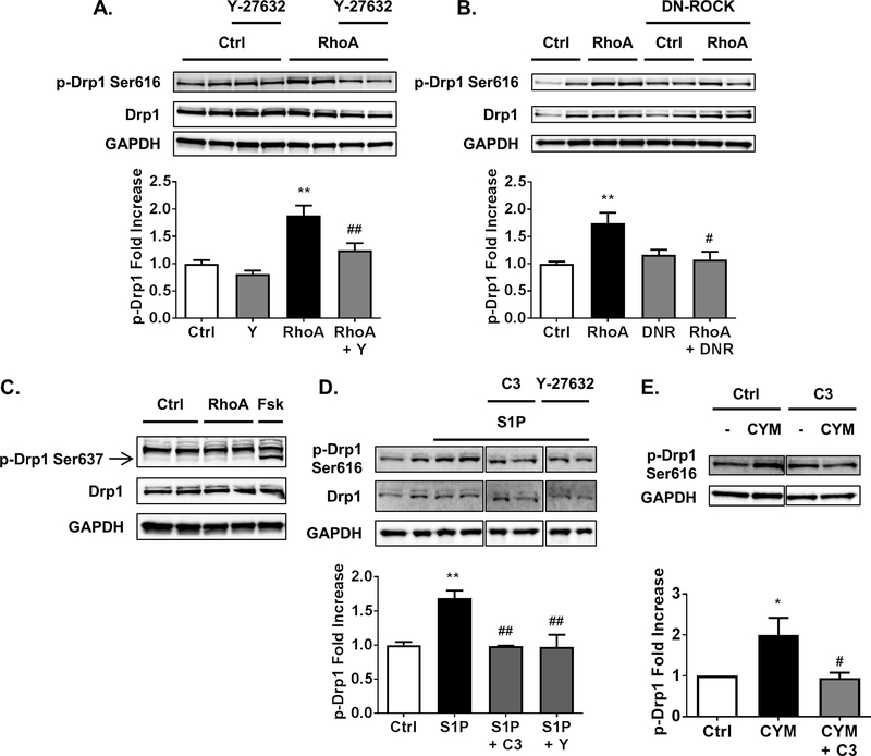
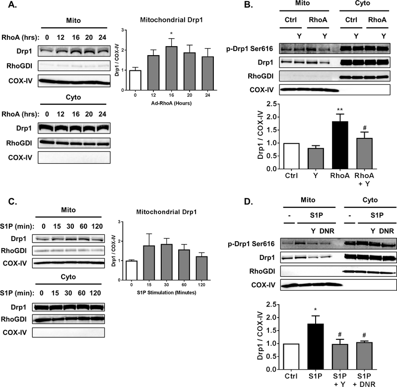
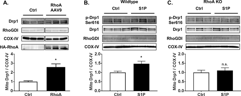
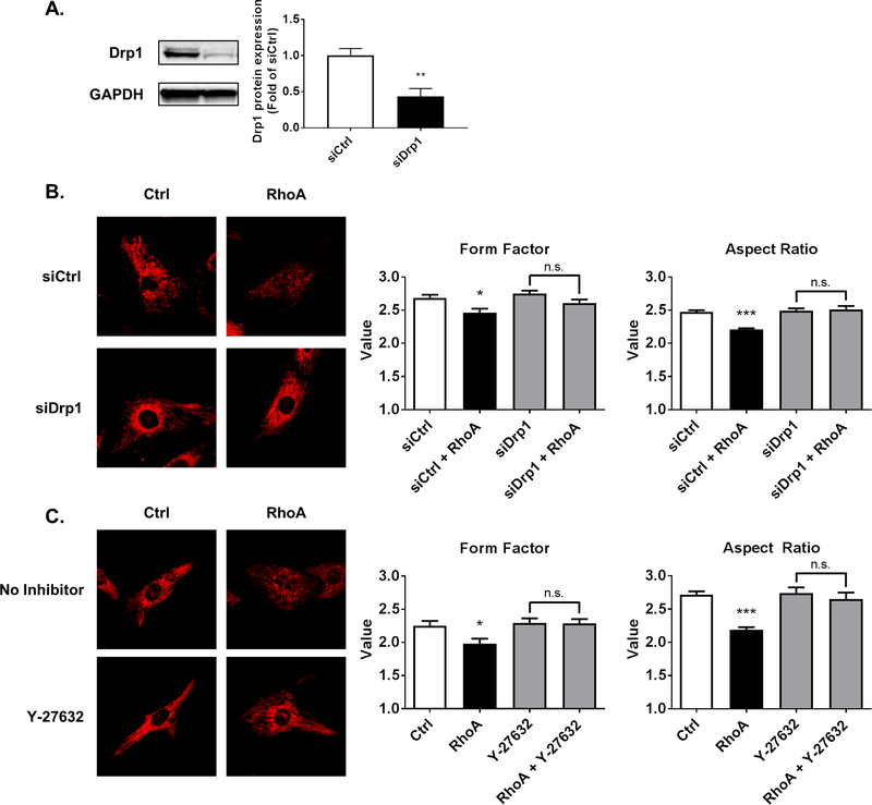
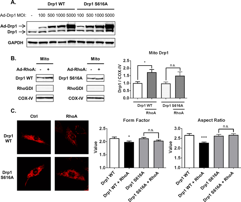
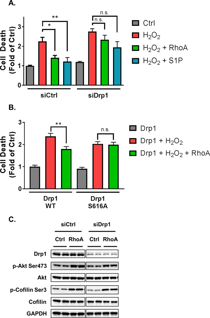
Cardiac ischemia/reperfusion, loss of blood flow and its subsequent restoration, causes damage to the heart. Oxidative stress from ischemia/reperfusion leads to dysfunction and death of cardiomyocytes, increasing the risk of progression to heart failure. Alterations in mitochondrial dynamics, in particular mitochondrial fission, have been suggested to play a role in cardioprotection from oxidative stress. We tested the hypothesis that activation of RhoA regulates mitochondrial fission in cardiomyocytes. Our studies show that expression of constitutively active RhoA in cardiomyocytes increases phosphorylation of Dynamin-related protein 1 (Drp1) at serine-616, and leads to localization of Drp1 at mitochondria. Both responses are blocked by inhibition of Rho-associated Protein Kinase (ROCK). Endogenous RhoA activation by the GPCR agonist sphingosine-1-phosphate (S1P) also increases Drp1 phosphorylation and its mitochondrial translocation in a RhoA and ROCK dependent manner. Consistent with the role of mitochondrial Drp1 in fission, RhoA activation in cardiomyocytes leads to formation of smaller mitochondria and this is attenuated by inhibition of ROCK, by siRNA knockdown of Drp1 or by expression of a phosphorylation-deficient Drp1 S616A mutant. In addition, activation of RhoA prevents cell death in cardiomyocytes challenged by oxidative stress and this protection is blocked by siRNA knockdown of Drp1 or by Drp1 S616A expression. Taken together our findings demonstrate that RhoA activation can regulate Drp1 to induce mitochondrial fission and subsequent cellular protection, implicating regulation of fission as a novel mechanism contributing to RhoA-mediated cardioprotection.

Keywords: Cardioprotection; Drp1; Fission; Mitochondria; RhoA; Sphingosine-1-phosphate.


Conflict of interest statement

Conflicts of interest

The authors declare that they have no conflicts of interest with the contents of this article.

Figures

Fig. 1.
Fig. 1.
RhoA activation through ROCK stimulates phosphorylation of Drp1 at serine-616. A, NRVMs adenovirally expressing activated RhoA or GFP control, in the absence or presence of 10 μM of ROCK inhibitor Y-27632. Whole cell lysates were analyzed by SDS-PAGE and Western blot for p-Drp1 at serine-616 and total Drp1 levels. **p < .01 vs. Ctrl, ##p < .01 vs. RhoA (n=5–6). B, NRVMs adenovirally expressing activated RhoA or GFP control, with and without co-expression of dominant-negative ROCK (DNR). Whole cell lysates were analyzed by SDS-PAGE and Western blot for p-Drp1 at serine-616 and total Drp1 levels. **p < .01 vs. Ctrl, #p < .05 vs. RhoA (n=4–5). C, A representative Western blot for p-Drp1 at serine-637 and total Drp1 in whole cell lysates from NRVMs adenovirally expressing activated RhoA or GFP control, or stimulated for 30 min with 10 μM forskolin (Fsk) as a positive control for PKA-dependent phosphorylation of Drp1 at serine-637. D, NRVMs were pretreated with C3 Rho inhibitor (1.5 μg/ml for 6 h pretreatment) or ROCK inhibitor Y-27632, then stimulated with 300 nM S1P for 30 min. Whole cell lysates were analyzed by SDS-PAGE and Western blot for p-Drp1 at serine-616 and total Drp1 levels. **p < .01 vs. Ctrl, ##p < .01 vs. S1P (n=4–6). E, NRVMs with or without pretreatment of C3 Rho inhibitor were stimulated by 10 μM of S1PR3 selective agonist CYM-51736 (CYM) for 30 min and cell lysates were subjected to Western blot for p-Drp1 at serine-616. *p < .05 vs. Ctrl, #p < .05 vs. CYM (n= 4).
Fig. 2.
Fig. 2.
RhoA activation through ROCK increases mitochondrial levels of Drp1 in cardiomyocytes. A, NRVMs adenovirally expressing activated RhoA for the amount of time indicated following infection. Mitochondrial and cytosolic fractions analyzed by SDS-PAGE and Western blot for Drp1 in respective fractions. COX-IV and RhoGDI were used as controls for loading of mitochondrial and cytosolic fractions respectively. *p < .05 vs. 0 h (n=5) B, NRVMs adenovirally expressing activated RhoA or GFP control, in the absence or presence of 10 μM of ROCK inhibitor Y-27632. Mitochondrial and cytosolic fractions analyzed by SDS-PAGE and Western blot for Drp1 in respective fractions. **p < .01 vs. Ctrl, #p < .05 vs. RhoA (n=5). C, NRVMs with stimulation by 300nM S1P for the amount of time indicated. Mitochondrial and cytosolic fractions analyzed by SDS-PAGE and Western blot for Drp1 in respective fractions. D, NRVMs in the absence or presence of 10 μM of ROCK inhibitor Y-27632 (Y) or adenovirally expressing dominant-negative ROCK (DNR), were stimulated by 300 nM S1P for 30 min. Mitochondrial and cytosolic fractions analyzed by SDS-PAGE and Western blot for Drp1 in respective fractions. **p < .05 vs. Ctrl, #p < .05 vs. S1P (n=4).
Fig. 3.
Fig. 3.
RhoA activation increases mitochondrial Drp1 levels in the adult mouse heart. A, Mice were injected with RhoA AAV9 or corresponding control for 2 weeks. Mitochondrial fractions of mouse hearts analyzed by SDS-PAGE and Western blot for Drp1. COX-IV and RhoGDI were used as mitochondrial and cytosolic markers respectively. *p < .05 vs. Ctrl (n=3). B, C, Mouse hearts from wildtype (B) or cardiac-specific RhoA knockouts (C) were subjected to ex vivo Langendorff perfusion with or without 300 nM S1P for 15 min. Mitochondrial fractions analyzed by SDS-PAGE and Western blot for Drp1. *p < .05 vs. Ctrl (n=7).
Fig. 4.
Fig. 4.
RhoA activation induces mitochondrial fission in cardiomyocytes through ROCK and Drp1. A, NRVMs were transfected with either control siRNA (siCtrl) or siRNA for Drp1 (siDrp1) for 48 h. **p < .01 vs. siCtrl (n=6). B, Confocal microscopy of NRVMs treated with either control or Drp1 siRNA for 48 h, and subsequent adenoviral expression of either activated RhoA or control. MitoTracker DeepRed (30 nM) was loaded into cardiomyocytes to visualize mitochondria. Particle analysis was carried out to calculate form factor and aspect ratio as indicators of circularity to assess mitochondrial size. *p < .05 vs. siCtrl, ***p < .001 vs. siCtrl (n=40). C, Confocal microscopy images of NRVMs with adenoviral expression of either activated RhoA or control, and absence or presence of ROCK inhibitor Y-27632 (10 μM). MitoTracker DeepRed was used to visualize mitochondria. Particle analysis was carried out to calculate form factor and aspect ratio as indicators of circularity to assess mitochondrial size. *p < .05 vs. Ctrl, ***p < .001 vs. Ctrl (n=30).
Fig. 5.
Fig. 5.
Phosphorylation of Drp1 at serine-616 is required for RhoA-induced mitochondrial fission. A, NRVMs were infected with adenovirus expressing wildtype or S616A phosphorylation-defective mutant Drp1. Whole cell lysates were analyzed by Western blot for Drp1. B, NRVMs were infected with 500 MOI of adenovirus expressing Drp1 wildtype or S616A, and active RhoA or GFP control were adenovirally co-expressed. Mitochondrial fractions were analyzed by Western blot for Drp1, with COX-IV and RhoGDI as mitochondrial and cytosolic markers respectively. *p < .05 vs. Ctrl (n=6). C, Fluorescent confocal microscopy images of NRVMs with adenoviral expression of wildtype or S616A phosphorylation-defective mutant Drp1, and adenoviral co-expression of activated RhoA or control, using MitoTracker DeepRed to visualize mitochondria. Particle analysis was carried out to calculate form factor and aspect ratio as indicators of circularity to assess mitochondrial size. *p < .05 vs. Drp1 WT, ***p < .001 vs. Drp1 WT (n=30).
Fig. 6.
Fig. 6.
Drp1 contributes to RhoA-mediated cardioprotection from oxidative stress. A, Following 48 h of control or Drp1 siRNA treatment, NRVMs were infected with adenovirus expressing activated RhoA or GFP control for 12 h and treated with or without S1P for 30 min. NRVMs were then treated with 50 μM H2O2 for 16 h. Cell death was quantified by Cell Death Detection ELISAPLUS POD assay of nucleus-free lysates. *p < .05 vs. H2O2, **p < .01 vs. H2O2 in control (n=6). B, NRVMs were infected with adenovirus expressing wildtype or S616A phosphorylation-defective mutant Drp1, and adenoviral co-expression of activated RhoA or GFP control for 24 h. NRVMs were then treated with 50 μM H2O2 for 16 h. Cell death was quantified by Cell Death Detection ELISAPLUS POD assay of nucleus-free lysates. **p < .01 vs. Drp1 + H2O2 (n=6). C, NRVMs were treated with control or Drp1 siRNA knockdown for 48 h. Subsequently, NRVMs were infected with adenovirus expressing activated RhoA or GFP control, and whole cell lysates were analyzed by Western blot for Drp1 to confirm knockdown, and of p-Akt, total Akt, p-cofilin, and total cofilin to assess phosphorylation of these proteins with and without Drp1 siRNA.

References

    1. Moe GW, Marin-Garcia J, Role of cell death in the progression of heart failure, Heart Fail. Rev 21 (2) (2016) 157–167. - PubMed
    1. Del Re DP, Miyamoto S, Brown JH, Focal adhesion kinase as a RhoA-activable signaling scaffold mediating Akt activation and cardiomyocyte protection, J. Biol. Chem 283 (51) (2008) 35622–35629. - PMC - PubMed
    1. Xiang SY, Vanhoutte D, Del Re DP, Purcell NH, Ling H, Banerjee I, Bossuyt J, Lang RA, Zheng Y, Matkovich SJ, Miyamoto S, Molkentin JD, Dorn GW 2nd, Brown JH, RhoA protects the mouse heart against ischemia/reperfusion injury, J. Clin. Invest 121 (8) (2011) 3269–3276. - PMC - PubMed
    1. Mozzicato S, Joshi BV, Jacobson KA, Liang BT, Role of direct RhoA-phospholipase D1 interaction in mediating adenosine-induced protection from cardiac ischemia, FASEB J. 18 (2) (2004) 406–408. - PubMed
    1. Krijnen PA, Sipkens JA, Molling JW, Rauwerda JA, Stehouwer CD, Muller A, Paulus WJ, van Nieuw Amerongen GP, Hack CE, Verhoeven AJ, van Hinsbergh VW, Niessen HW, Inhibition of Rho-ROCK signaling induces apoptotic and non-apoptotic PS exposure in cardiomyocytes via inhibition of flippase, J. Mol. Cell. Cardiol 49 (5) (2010) 781–790. - PubMed

MeSH terms