Review
. 2018 Jun 27;19(7):1886.
doi: 10.3390/ijms19071886.

GPCRs in Cancer: Protease-Activated Receptors, Endocytic Adaptors and Signaling

Affiliations
Review

GPCRs in Cancer: Protease-Activated Receptors, Endocytic Adaptors and Signaling

Aleena K S Arakaki et al. Int J Mol Sci. .

Abstract

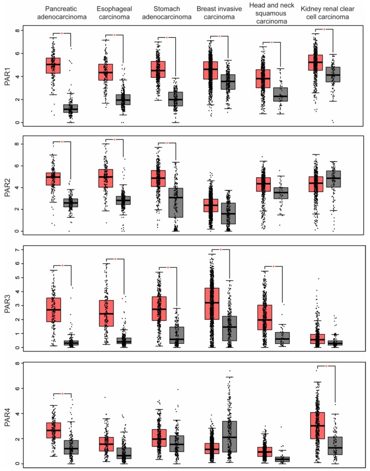
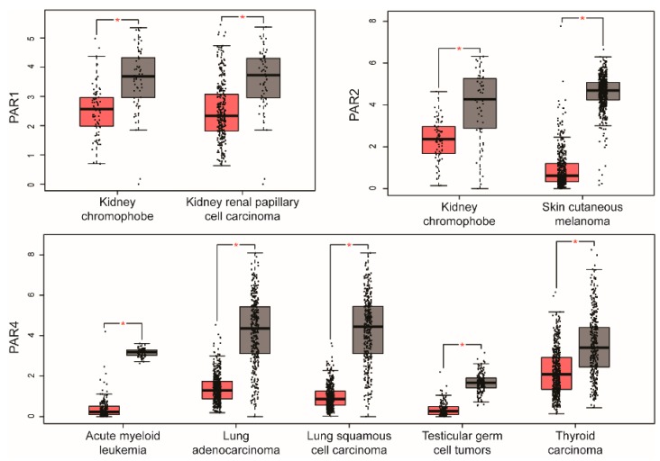
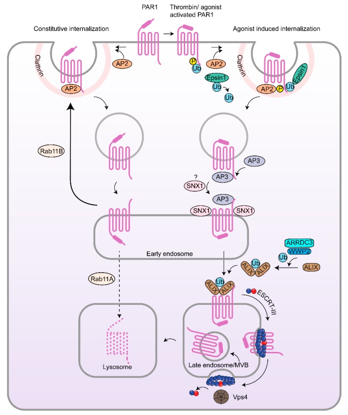
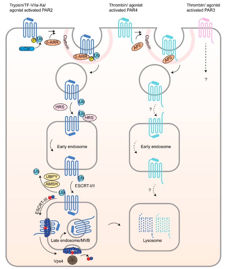
G protein-coupled receptors (GPCRs) are a large diverse family of cell surface signaling receptors implicated in various types of cancers. Several studies indicate that GPCRs control many aspects of cancer progression including tumor growth, invasion, migration, survival and metastasis. While it is known that GPCR activity can be altered in cancer through aberrant overexpression, gain-of-function activating mutations, and increased production and secretion of agonists, the precise mechanisms of how GPCRs contribute to cancer progression remains elusive. Protease-activated receptors (PARs) are a unique class of GPCRs implicated in cancer. PARs are a subfamily of GPCRs comprised of four members that are irreversibly activated by proteolytic cleavage induced by various proteases generated in the tumor microenvironment. Given the unusual proteolytic irreversible activation of PARs, expression of receptors at the cell surface is a key feature that influences signaling responses and is exquisitely controlled by endocytic adaptor proteins. Here, we discuss new survey data from the Cancer Genome Atlas and the Genotype-Tissue Expression projects analysis of expression of all PAR family member expression in human tumor samples as well as the role and function of the endocytic sorting machinery that controls PAR expression and signaling of PARs in normal cells and in cancer.

Keywords: ARRDC3; arrestins; breast cancer; invasion; lysosomes; metastasis.


Conflict of interest statement

This manuscript reports unpublished work that is not under consideration for publication elsewhere. All co-authors on this work agree to its content and hereby declare no competing commercial interests relating to this submitted work.

Figures

Figure 1
Figure 1
Co-expression of protease-activated receptors in human cancers. GEPIA analysis revealed that multiple PARs are significantly overexpressed in various cancer types. Pancreatic adenocarcinoma (179 tumor, 171 normal) overexpresses all PARs compared to normal tissue; esophageal carcinoma (182 tumor, 286 normal) and stomach adenocarcinoma (408 tumor, 211 normal) overexpress PAR1, PAR2, and PAR3, whereas breast invasive carcinoma (1085 tumor, 291 normal) and head and neck squamous carcinoma (519 tumor, 44 normal) overexpress PAR1 and PAR3 and kidney renal clear cell carcinoma (523 tumor, 100 normal) overexpress PAR1 and PAR4. The RNA-seq data are expressed as relative gene expression using transformed log2 (TPM+1) value (Y-axis) of tumor (red) and normal (grey) samples from different cancer types and displayed as a whisker plot. The whisker plot solid horizontal black line is the median, the box represents the upper and lower quartiles and the two lines (whiskers) outside the box extend to the highest and lowest observations of the sample population. The difference in PAR expression in tumors compared to normal tissue control is significant based on one-way ANOVA (* p < 0.01). TPM, transcript per million.
Figure 2
Figure 2
Human cancers with significant PAR1 and PAR2 overexpression. A survey of cancers using GEPIA analysis revealed several cancer types with only PAR1 and PAR2 overexpression compared to normal tissue, including colon adenocarcinoma (275 tumor, 349 normal), glioblastoma multiforme (163 tumor, 207 normal), ovarian serous cystadenocarcinoma (426 tumor, 88 normal), and rectum adenocarcinoma (92 tumor, 318 normal). The RNA-seq data are expressed as the relative gene expression using transformed log2 (TPM+1) value (Y-axis) of tumor (red) and normal (grey) in different cancer types and displayed as whisker plots as described in Figure 1. The data showed a significant difference in PAR1 and PAR2 expression in tumors compared to normal tissue using one-way ANOVA (* p < 0.01).
Figure 3
Figure 3
Human cancers with downregulation of PAR1, PAR2 and PAR4 expression. A few cancers displayed significant decreases in expression of PARs based on GEPIA analysis. PAR1 showed significantly downregulation in kidney chromophobe (66 tumor, 53 normal) and kidney renal papillary cell carcinoma (286 tumor, 60 normal). PAR2 was significantly downregulated in kidney chromophobe and skin cutaneous melanoma (461 tumor, 558 normal). PAR4 was significantly downregulated in acute myeloid leukemia (173 tumor, 70 normal), lung adenocarcinoma (483 tumor, 347 normal), lung squamous cell carcinoma (486 tumor, 338 normal), testicular germ cell tumors (137 tumor, 165 normal), and thyroid carcinoma (512 tumor, 337 normal). RNA-seq data are expressed as relative gene expression using transformed log2 (TPM+1) value (Y-axis) of tumor (red) and normal (grey) samples from different cancer types and displayed as whisker plots as shown in Figure 1. The data showing differences in PAR expression in tumor compared to normal tissue are significant based on one-way ANOVA (* p < 0.01).
Figure 4
Figure 4
Endocytic trafficking of PAR1. PAR1 undergoes constitutive and agonist-activated internalization induced by thrombin cleavage of the N-terminus or by the peptide agonist SFLLRN. Unactivated PAR1 is constitutively internalized by AP-2 recognition of a distal tyrosine-based motif within the cytoplasmic C-terminus of PAR1. Internalized PAR1 is then sorted to early endosomes and then recycled back to the cell surface via a Rab11B-dependent pathway, whereas a small pool of receptor escapes recycling and is sorted by Rab11A to lysosomes and degraded. In contrast, agonist activation of PAR1 results in rapid phosphorylation and ubiquitination and internalization through a dynamin- and clathrin-dependent pathway mediated by AP-2 and epsin-1. AP-2 binds the phosphorylated distal C-terminus of activated PAR1 rather than the tyrosine-based motif to regulate activated PAR1 internalization. PAR1 activation also promotes epsin-1 deubiquitination, facilitating the ability of epsin-1 to bind activated PAR1 to facilitate internalization. Internalized PAR1 is then sorted sequentially at early endosomes by engaging AP-3 and SNX1 followed by ALIX, which requires ARRDC3 and WWP2-mediated ALIX ubiquitination and dimerization. ALIX, ARRDC3 and WWP2 are essential for targeting PAR1 to intraluminal vesicles of multivesicular bodies (MVBs)/late endosomes via ECSRT-III charged MVB protein 4 (CHMP4) and AAA-ATPase vacuolar protein sorting 4 (Vps4). Degradation of PAR1 in lysosomes is ultimately required for signal termination.
Figure 5
Figure 5
Endocytic trafficking of PAR2, PAR3 and PAR4. Agonist-activation of PAR2 can occur by trypsin, tissue factor (TF)-VIIa-Xa or peptide agonist SLIGKV and induces phosphorylation and ubiquitination followed by receptor internalization, which is mediated by β-arrestins (β-ARR) through a clathrin-dependent pathway. Ubiquitination of activated PAR2 by the E3 ubiquitin ligase c-Cbl, functions primarily in PAR2 endosomal-lysosomal trafficking. Ubiquitinated PAR2 is recognized by HRS at early endosomes and required for lysosomal sorting, after deubiquitination mediated by the AMSH and UBPY deubiquitinating enzymes that enable sorting of PAR2 to MVBs by the ESCRT-III/Vps4 machinery and ultimately degradation. Agonist-activation of PAR4 by thrombin or AYPGKF induces internalization mediated by AP-2 and occurs through a clathrin-dependent pathway similar to PAR1. AP-2 binds activated PAR4 by recognizing a tyrosine-based sorting motif present in the third intracellular loop of the receptor. Internalized PAR4 is then sorted to endosomes and lysosomes for degradation. The endocytic adaptor proteins that facilitate PAR4 endosomal-lysosomal trafficking are unknown. The mechanisms that control thrombin-activated PAR3 internalization and trafficking to lysosomes is not known.

References

    1. Singh A., Nunes J.J., Ateeq B. Role and therapeutic potential of g-protein coupled receptors in breast cancer progression and metastases. Eur. J. Pharmacol. 2015;763:178–183. doi: 10.1016/j.ejphar.2015.05.011. - DOI - PMC - PubMed
    1. Bar-Shavit R., Maoz M., Kancharla A., Nag J.K., Agranovich D., Grisaru-Granovsky S., Uziely B. G protein-coupled receptors in cancer. Int. J. Mol. Sci. 2016;17:1320. doi: 10.3390/ijms17081320. - DOI - PMC - PubMed
    1. O’Hayre M., Vazquez-Prado J., Kufareva I., Stawiski E.W., Handel T.M., Seshagiri S., Gutkind J.S. The emerging mutational landscape of g proteins and g-protein-coupled receptors in cancer. Nat. Rev. Cancer. 2013;13:412–424. doi: 10.1038/nrc3521. - DOI - PMC - PubMed
    1. Santos R., Ursu O., Gaulton A., Bento A.P., Donadi R.S., Bologa C.G., Karlsson A., Al-Lazikani B., Hersey A., Oprea T.I., et al. A comprehensive map of molecular drug targets. Nat. Rev. Drug Discov. 2017;16:19–34. doi: 10.1038/nrd.2016.230. - DOI - PMC - PubMed
    1. Sriram K., Insel P.A. Gpcrs as targets for approved drugs: How many targets and how many drugs? Mol. Pharmacol. 2018 doi: 10.1124/mol.117.111062. - DOI - PMC - PubMed

MeSH terms

Substances