. 2018 Jul;27(7):1080-1095.
doi: 10.1177/0963689718780942. Epub 2018 Jun 28.

Mesenchymal Stem Cell-Derived Extracellular Vesicles Improve the Renal Microvasculature in Metabolic Renovascular Disease in Swine

Affiliations

Mesenchymal Stem Cell-Derived Extracellular Vesicles Improve the Renal Microvasculature in Metabolic Renovascular Disease in Swine

Alfonso Eirin et al. Cell Transplant. 2018 Jul.

Abstract

Background: Extracellular vesicles (EVs) released from mesenchymal stem/stromal cells (MSCs) mediate their paracrine effect, but their efficacy to protect the microcirculation of the kidney is unknown. Using a novel swine model of unilateral renovascular disease (RVD) complicated by metabolic syndrome (MetS), we tested the hypothesis that EVs would attenuate renal microvascular loss.

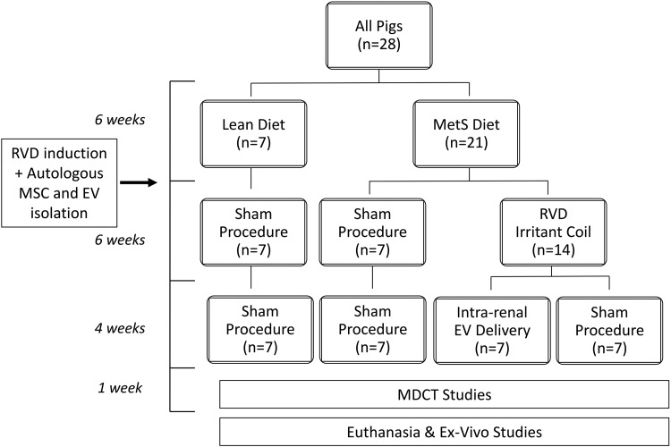
Methods: Four groups of pigs ( n = 7 each) were studied after 16 weeks of diet-induced MetS and RVD (MetS+RVD), MetS+RVD treated 4 weeks earlier with a single intra-renal delivery of EVs harvested from autologous adipose tissue-derived MSCs, and Lean and MetS Sham controls. Stenotic-kidney renal blood flow (RBF) and glomerular filtration rate (GFR) were measured in-vivo (fast CT), whereas EV characteristics, renal microvascular architecture (micro-CT), and injury pathways were studied ex-vivo.

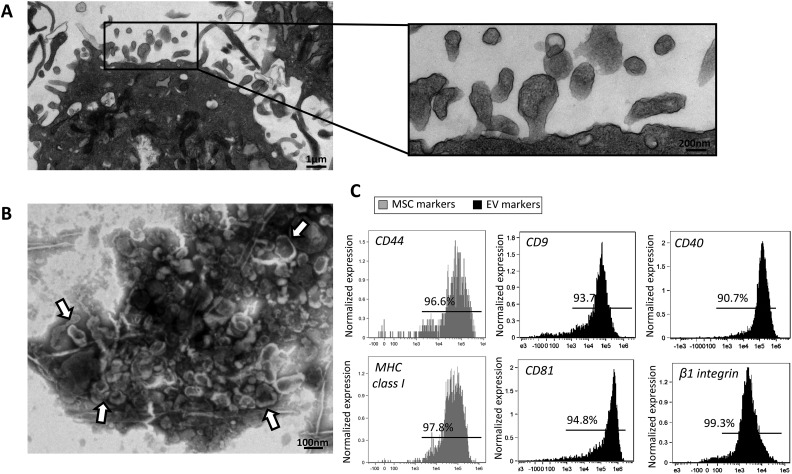
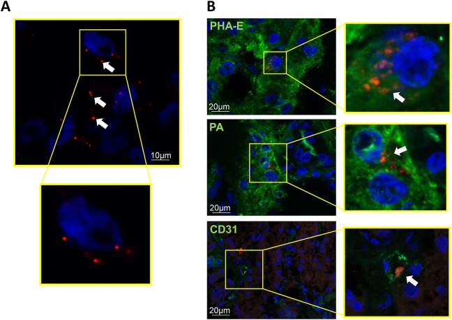
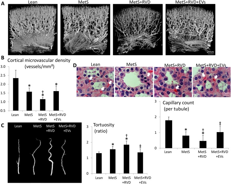
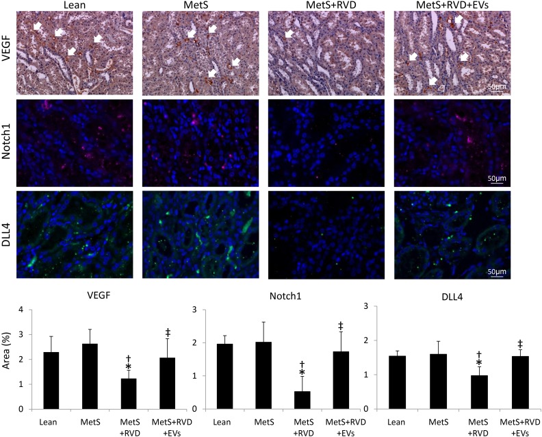
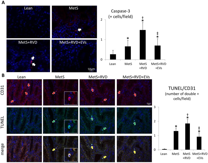
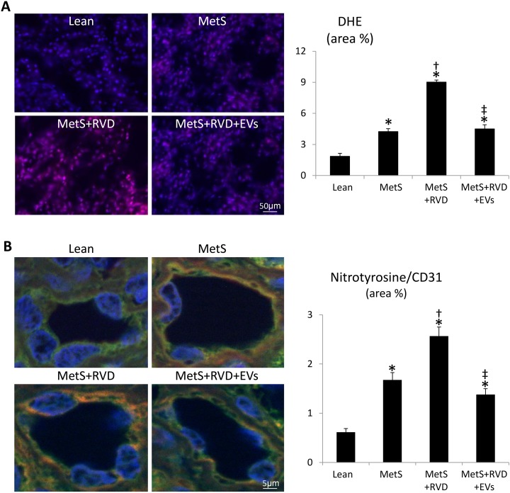
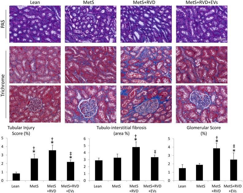
Results: mRNA sequencing and proteomic analysis revealed that EVs are packed with several pro-angiogenic genes and proteins, such as vascular endothelial growth factor. Labeled EVs were detected in the stenotic kidney 4 weeks after injection internalized by tubular and endothelial cells. EVs restored renal expression of angiogenic factors and improved cortical microvascular and peritubular capillary density. Renal apoptosis, oxidative stress, tubular injury, and fibrosis were also attenuated in EV-treated pigs. RBF and GFR decreased in MetS+RVD compared with MetS, but normalized in MetS+RVD+EVs.

Conclusions: Intra-renal delivery of MSC-derived EVs bearing pro-angiogenic properties restored the renal microcirculation and in turn hemodynamics and function in chronic experimental MetS+RVD. Our study suggests a novel therapeutic potential for MSC-derived EVs in restoring renal hemodynamics in experimental MetS+RVD.

Keywords: extracellular vesicles; mesenchymal stem cells; metabolic syndrome; microcirculation; renovascular disease.


Conflict of interest statement

Declaration of Conflicting Interests: The authors declared no potential conflicts of interest with respect to the research, authorship, and/or publication of this article.

Figures

Figure 1.
Figure 1.
Schematic of the experimental protocol. At baseline, pigs were fed either a MetS (n = 21) or Lean diet (n = 7). Six weeks later, RVD was induced in 14 MetS pigs, whereas 7 Lean and 7 MetS pigs underwent a sham procedure. Six weeks after induction of RVD, MetS+RVD pigs received a single intra-renal infusion of either autologous MSC-derived EVs or vehicle (n = 7 each). Other MetS and Lean pigs underwent sham procedures (n = 7 each). Four weeks later, pigs were studied in-vivo and ex-vivo.
Figure 2.
Figure 2.
A: Transmission electron microscopy showing EVs released from MSCs. B: EVs exhibit a classic morphology on negative staining. C: Fluorescence-activated cell sorting reveals that EVs express common EV and MSC markers.
Figure 3.
Figure 3.
A: EV clusters were detected in the swine kidney 4 weeks after intra-renal delivery (arrows). B: Immunofluorescence co-staining with Phaseolus vulgaris erythroagglutinin (PHA-E), peanut agglutinin (PA), and CD31, shows EV engraftment in proximal and distal tubules, and endothelial cells, respectively.
Figure 4.
Figure 4.
EVs improve microvascular architecture in MetS+RVD. A: Representative 3D micro-computed tomography images of the pig kidney showing improved microvascular architecture in EV-treated pigs. B: Quantification of spatial density of renal cortical microvessels (left) and microvascular tortuosity (right). D: Representative renal hemotoxylin and eosin (H&E) staining and quantification of peritubular capillary density. *p < 0.05 vs. Lean; p < 0.05 vs. MetS; p < 0.05 vs. MetS+RVD.
Figure 5.
Figure 5.
EVs improve angiogenic signaling in MetS+RVD. Representative stenotic kidney staining (×40) for the pro-angiogenic factors VEGF, Notch-1, and Notch ligand delta-like-4 (DLL4), and their quantification. *p < 0.05 vs. Lean; p < 0.05 vs. MetS; p < 0.05 vs. MetS+RVD.
Figure 6.
Figure 6.
EVs decreased endothelial cell apoptosis in MetS+RVD. A: Fluorescent renal staining (40×) and quantification of caspase-3. B: Double renal fluorescence staining with terminal deoxynucleotidyl transferase-mediated dUTP nick end labeling (TUNEL, green arrows) and CD31 (green arrows). Yellow arrows indicated double + cells. *p < 0.05 vs. Lean; p < 0.05 vs. MetS; p < 0.05 vs. MetS+RVD.
Figure 7.
Figure 7.
EVs decreased vascular oxidative stress in MetS+RVD. A: Fluorescent renal staining (40×) and quantification of dihydroethidium (DHE). Double renal fluorescence staining with nitrotyrosine (red arrows) and CD31 (green arrows) shows endothelial-cell-specific oxidative stress. Yellow arrows indicate double + cells. *p < 0.05 vs. Lean; p < 0.05 vs. MetS; p < 0.05 vs. MetS+RVD.
Figure 8.
Figure 8.
Representative kidney periodic acid-Schiff (PAS) and trichrome staining (40×), and quantifications of tubular injury, tubulo-interstitial fibrosis, and glomerular score. *p < 0.05 vs. Lean; p < 0.05 vs. MetS; p < 0.05 vs. MetS+RVD.

References

    1. Martins D, Ani C, Pan D, Ogunyemi O, Norris K. Renal dysfunction, metabolic syndrome and cardiovascular disease mortality. J Nutr Metab. 2010;2010:1–8. - PMC - PubMed
    1. Ferraro PM, Lupo A, Yabarek T, Graziani MS, Bonfante L, Abaterusso C, Gambaro G, Incipe Study G. Metabolic syndrome, cardiovascular disease, and risk for chronic kidney disease in an italian cohort: Analysis of the incipe study. Metab Syndr Relat Disord. 2011;9(5):381–388. - PubMed
    1. Lerman L, Textor SC. Pathophysiology of ischemic nephropathy. Urol Clin North Am. 2001;28(4):793–803, ix. - PubMed
    1. Davies MG, Saad WE, Bismuth J, Naoum JJ, Peden EK, Lumsden AB. Impact of metabolic syndrome on the outcomes of percutaneous renal angioplasty and stenting. J Vasc Surg. 2010;51(4):926–932. - PubMed
    1. Zou X, Gu D, Xing X, Cheng Z, Gong D, Zhang G, Zhu Y. Human mesenchymal stromal cell-derived extracellular vesicles alleviate renal ischemic reperfusion injury and enhance angiogenesis in rats. Am J Transl Res. 2016;8(10):4289–4299. - PMC - PubMed

Publication types