Case Reports
. 2018 Jun 28;13(1):82.
doi: 10.1186/s13019-018-0764-z.

Successful emergent repair of a subacute left ventricular free wall rupture after acute inferoposterolateral myocardial infarction

Affiliations
Case Reports

Successful emergent repair of a subacute left ventricular free wall rupture after acute inferoposterolateral myocardial infarction

Arjan J F P Verhaegh et al. J Cardiothorac Surg. .

Abstract

Background: Myocardial rupture is an important and catastrophic complication of acute myocardial infarction. A dramatic form of this complication is a left ventricular free wall rupture (LVFWR).

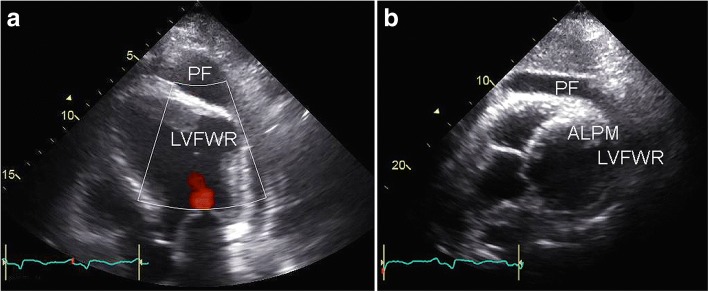
Case presentation: A 70-year-old man with acute inferoposterolateral myocardial infarction and single-vessel coronary artery disease underwent emergency percutaneous coronary intervention (PCI). The circumflex coronary artery was successfully stented with a drug-eluting stent. Fifty days after PCI the patient experienced progressive fatigue and chest pain with haemodynamic instability. Transthoracic echocardiography showed a covered LVFWR of the lateral wall. The patient underwent successful emergent surgical repair of the LVFWR.

Conclusions: In the current era of swift PCI, mechanical complications of acute myocardial infarction, such as LVFWR, are rare. The consequences, however, are haemodynamic deterioration and imminent death. This rare diagnosis should always be considered when new cardiovascular symptoms or haemodynamic instability develop after myocardial infarction, even beyond one month after the initial event. Timely diagnosis and emergency surgery are required for successful treatment of this devastating complication.

Keywords: Myocardial infarction, heart rupture, percutaneous coronary intervention, cardiac surgical procedures.


Conflict of interest statement

Consent for publication

Informed consent was obtained from the patient for publication of this case report and any accompanying images.

Competing interests

The authors declare that they have no competing interests.

Publisher’s Note

Springer Nature remains neutral with regard to jurisdictional claims in published maps and institutional affiliations.

Figures

Fig. 1
Fig. 1
Preoperative transthoracic echocardiographic imaging. Preoperative transthoracic echocardiography showing covered left ventricular free wall rupture (LVFWR) of the lateral wall in the apical four-chamber view (a) and the parasternal short-axis view (b). Note extensive pericardial fluid (PF) and the close proximity of the rupture to the anterolateral papillary muscle (ALPM)
Fig. 2
Fig. 2
Intraoperative photographs. After carefully opening the chest and pericardium, cardiopulmonary bypass was instituted. Subsequently, cardioplegic cardiac arrest was achieved and the covered left ventricular free wall rupture of the lateral wall was visualized after careful manupulation of the heart (a, arrows). The left ventricular free wall rupture was only covered by a thin layer of epicardium. Opening of the thin-walled epicardial layer revealed the close proximity of the rupture to the anterolateral papillary muscle of the mitral valve (b). The defect was approximately 5–6 cm in diameter. The LVFWR was repaired by (1) approximation of the defect with a circumferential purse-string suture (a so-called “Fontan stitch”) (c, arrow), which reduced the diameter of the defect to 3 cm, (2) securing a Dacron patch on the endocardial surface of the heart (carefully avoiding sutures too close to the ALPM) (d, e) and (3) subsequently closing the ventriculotomy in two rows (a deep row with horizontal interrupted mattress sutures (f) and a superficial row with a continuous suture over a double layer of felt (g))

Similar articles

Cited by

References

    1. Reddy SG, Roberts WC. Frequency of rupture of the left ventricular free wall or ventricular septum among necropsy cases of fatal acute myocardial infarction since introduction of coronary care units. Am J Cardiol. 1989;63(13):906–911. doi: 10.1016/0002-9149(89)90137-9. - DOI - PubMed
    1. Yip HK, Wu CJ, Chang HW, Wang CP, Cheng CI, Chua S, et al. Cardiac rupture complicating acute myocardial infarction in the direct percutaneous coronary intervention reperfusion era. Chest. 2003;124(2):565–571. doi: 10.1378/chest.124.2.565. - DOI - PubMed
    1. Ibanez B, James S, Agewall S, Antunes MJ, Bucciarelli-Ducci C, Bueno H, et al. 2017 ESC guidelines for the management of acute myocardial infarction in patients presenting with ST-segment elevation. Web addenda. Eur Heart J. 2018;39(2):119–177. doi: 10.1093/eurheartj/ehx393. - DOI - PubMed
    1. Hutchins KD, Skurnick J, Lavenhar M, Natarajan GA. Cardiac rupture in acute myocardial infarction: a reassessment. Am J Forensic Med Pathol. 2002;23(1):78–82. doi: 10.1097/00000433-200203000-00017. - DOI - PubMed
    1. Batts KP, Ackermann DM, Edwards WD. Postinfarction rupture of the left ventricular free wall: clinicopathologic correlates in 100 consecutive autopsy cases. Hum Pathol. 1990;21(5):530–535. doi: 10.1016/0046-8177(90)90010-3. - DOI - PubMed

Publication types