. 2018 Jun 28;6(1):51.
doi: 10.1186/s40478-018-0553-x.

Non-inflammatory tumor microenvironment of diffuse intrinsic pontine glioma

Affiliations

Non-inflammatory tumor microenvironment of diffuse intrinsic pontine glioma

Grant L Lin et al. Acta Neuropathol Commun. .

Abstract

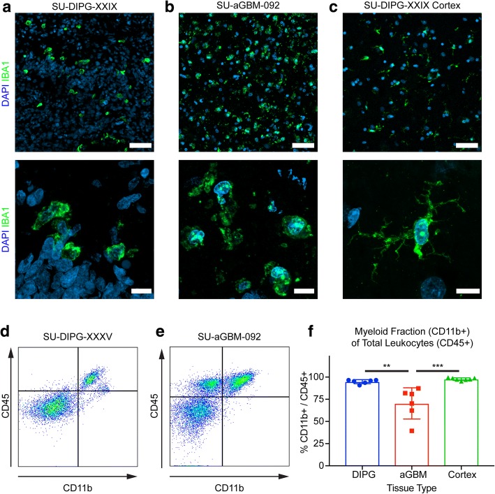
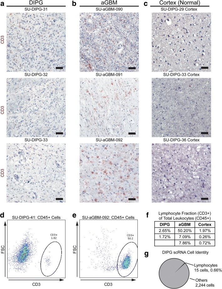
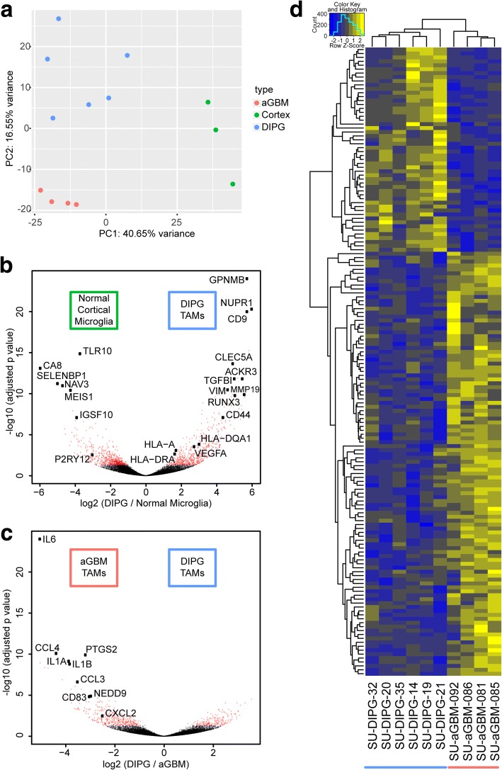
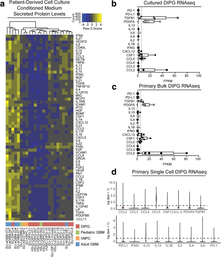
Diffuse intrinsic pontine glioma (DIPG) is a universally fatal malignancy of the childhood central nervous system, with a median overall survival of 9-11 months. We have previously shown that primary DIPG tissue contains numerous tumor-associated macrophages, and substantial work has demonstrated a significant pathological role for adult glioma-associated macrophages. However, work over the past decade has highlighted many molecular and genomic differences between pediatric and adult high-grade gliomas. Thus, we directly compared inflammatory characteristics of DIPG and adult glioblastoma (GBM). We found that the leukocyte (CD45+) compartment in primary DIPG tissue samples is predominantly composed of CD11b + macrophages, with very few CD3+ T-lymphocytes. In contrast, T-lymphocytes are more abundant in adult GBM tissue samples. RNA sequencing of macrophages isolated from primary tumor samples revealed that DIPG- and adult GBM-associated macrophages both express gene programs related to ECM remodeling and angiogenesis, but DIPG-associated macrophages express substantially fewer inflammatory factors than their adult GBM counterparts. Examining the secretome of glioma cells, we found that patient-derived DIPG cell cultures secrete markedly fewer cytokines and chemokines than patient-derived adult GBM cultures. Concordantly, bulk and single-cell RNA sequencing data indicates low to absent expression of chemokines and cytokines in DIPG. Together, these observations suggest that the inflammatory milieu of the DIPG tumor microenvironment is fundamentally different than adult GBM. The low intrinsic inflammatory signature of DIPG cells may contribute to the lack of lymphocytes and non-inflammatory phenotype of DIPG-associated microglia/macrophages. Understanding the glioma subtype-specific inflammatory milieu may inform the design and application of immunotherapy-based treatments.

Keywords: Diffuse intrinsic pontine glioma; Glioblastoma; Glioma; Immune microenvironment; Tumor-associated macrophage; Tumor-infiltrating lymphocyte.


Conflict of interest statement

Ethics approval and consent to participate

No animals were used in this study. All procedures performed in studies involving human participants were in accordance with the ethical standards of the institutional and/or national research committee and with the 1964 Helsinki declaration and its later amendments or comparable ethical standards.

Consent for publication

Informed consent was obtained from all individual participants included in the study prior to any tissue collection.

Competing interests

The authors declare that they have no competing interests.

Publisher’s Note

Springer Nature remains neutral with regard to jurisdictional claims in published maps and institutional affiliations.

Figures

Fig. 1
Fig. 1
DIPG and aGBM tumor-associated microglia/macrophages (a-c) Immunofluorescence of primary DIPG tumor tissue (a), primary adult GBM tumor tissue (b), and primary pediatric cerebral cortex (c) for the myeloid cell marker IBA1 (green) and DAPI counterstain (blue). The tumor-associated macrophages in a-b exhibit an amoeboid morphology with shorter processes and enlarged cell bodies, while the normal cortical microglia in c demonstrates the ramified morphology typical of "resting" microglia. Top scale bar = 50μm; bottom scale bar = 10μm (d-e) Representative FACS plots showing CD11b expression against CD45 expression of primary DIPG (d) and aGBM (e) tissue. Samples were gated on size, singularity, and viability prior to these plots (f) Quantification of myeloid fraction (CD11b+) of total leukocytes (CD45+) as calculated by flow cytometry in DIPG, aGBM, and normal cortex primary tissue. ** p < 0.005, *** p < 0.0005 by t-test with Tukey multiple comparison correction
Fig. 2
Fig. 2
DIPG and aGBM demonstrate differential CD3+ lymphocyte infiltration (a-c) Immunohistochemical staining for CD3 (red) of primary DIPG (a), adult GBM (b), and pediatric cerebral cortex (c) tissue, counterstained with hematoxylin (blue). Scale bar = 50 μm (d-e) FACS plots showing different CD3+ composition of CD45+ cells in DIPG (d) and aGBM (e). Samples were gated for size, singularity, viability, and CD45 positivity prior to these plots (f) Table of lymphocyte (CD3+) fraction of total leukocytes (CD45+) in primary DIPG, aGBM, and pediatric cortical tissue samples as calculated via flow cytometry of primary dissociated early autopsy samples. g Lymphocyte fraction of all sampled cells from primary single-cell RNA sequencing of DIPG diagnostic biopsy samples [7]
Fig. 3
Fig. 3
DIPG- and aGBM-associated macrophages exhibit distinct gene expression profiles (a) Principal components analysis of the top 500 varying genes across all samples demonstrates clusters corresponding to normal cortical microglia (green), DIPG-associated macrophages (blue), and aGBM-associated macrophages (red). b-c Volcano plot of log-fold change against adjusted p value for genes between normal cortical microglia and DIPG-associated macrophages (b), and between DIPG-associated macrophages and aGBM-associated macrophages (c). Red dots represent adjusted p value < 0.05, and selected significantly differentially expressed genes are identified (d) Heat map of normalized count values for differentially expressed genes between DIPG- and aGBM-associated macrophages. Hierarchical clustering demonstrates a distinct difference between the two groups
Fig. 4
Fig. 4
Patient-derived DIPG and aGBM cell cultures exhibit distinct cytokine secretion profiles (a) Hierarchical clustering of mean fluorescence intensity values of secreted factors in conditioned medium derived from patient-derived DIPG (red), aGBM (blue), pediatric GBM (green), and human neural precursor cell (hNPC) cultures (orange). Each column represents the average of samples tested in triplicate, and each row represents a separate measured cytokine (b-c) Box and whisker plot of FPKMs of cytokines and chemokines from patient-derived DIPG cell cultures (b) and primary bulk DIPG tissue (c) RNA sequencing data [11, 30, 35]. Dashed line represents FPKM = 5 (d) Violin plots of gene expression from single-cell RNA sequencing of diagnostic DIPG biopsy samples [7]. Dashed line represents log(tpm + 1) = 1

References

    1. Aguzzi A, Barres BA, Bennett ML. Microglia: scapegoat, saboteur, or something else? Science. 2013;339:156–161. doi: 10.1126/science.1227901. - DOI - PMC - PubMed
    1. Buczkowicz P, Hoeman C, Rakopoulos P, Pajovic S, Letourneau L, et al. Genomic analysis of diffuse intrinsic pontine gliomas identifies three molecular subgroups and recurrent activating. Nat Genet. 2014;46:451–456. doi: 10.1038/ng.2936. - DOI - PMC - PubMed
    1. Butowski N, Colman H, De Groot JF, Omuro AM, Nayak L, et al. Orally administered colony stimulating factor 1 receptor inhibitor PLX3397 in recurrent glioblastoma: an ivy Foundation early phase clinical trials consortium phase II study. Neuro-Oncology. 2016;18:557–564. doi: 10.1093/neuonc/nov245. - DOI - PMC - PubMed
    1. Cage TA, Samagh SP, Mueller S, Nicolaides T, Haas-Kogan D, et al. Feasibility, safety, and indications for surgical biopsy of intrinsic brainstem tumors in children. Childs Nerv Syst. 2013;29:1313–1319. doi: 10.1007/s00381-013-2101-0. - DOI - PubMed
    1. Cannarile MA, Weisser M, Jacob W, Jegg A-M, Ries CH, et al. Colony-stimulating factor 1 receptor (CSF1R) inhibitors in cancer therapy. J Immunotherapy Cancer. 2017;5:53. doi: 10.1186/s40425-017-0257-y. - DOI - PMC - PubMed

Publication types

MeSH terms