. 2018 Aug 17;293(33):12962-12974.
doi: 10.1074/jbc.RA118.003424. Epub 2018 Jun 28.

Pore properties of Orai1 calcium channel dimers and their activation by the STIM1 ER calcium sensor

Affiliations

Pore properties of Orai1 calcium channel dimers and their activation by the STIM1 ER calcium sensor

Xiangyu Cai et al. J Biol Chem. .

Abstract

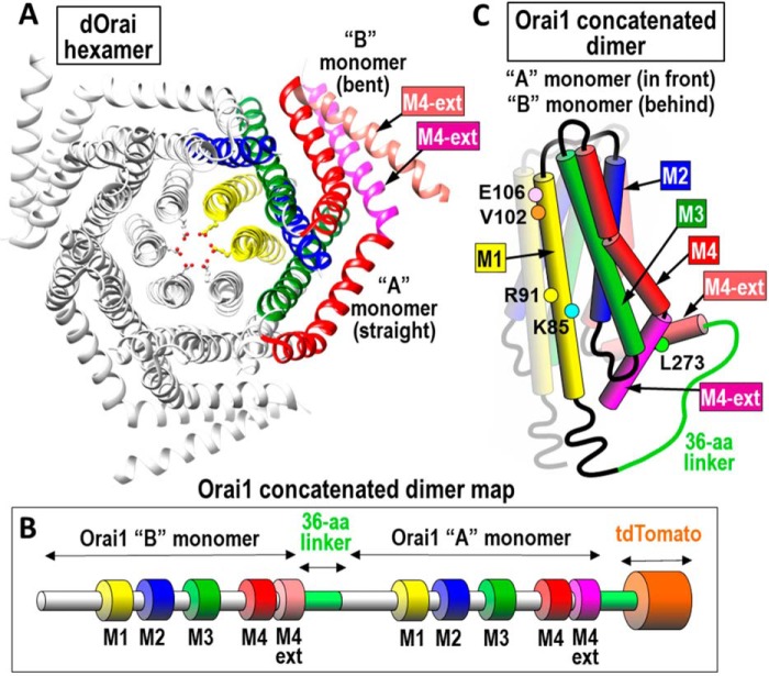
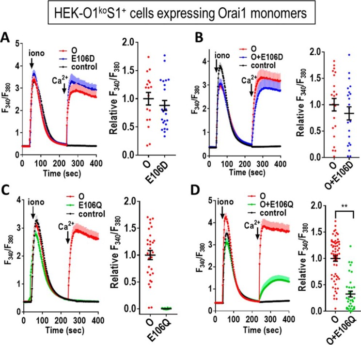
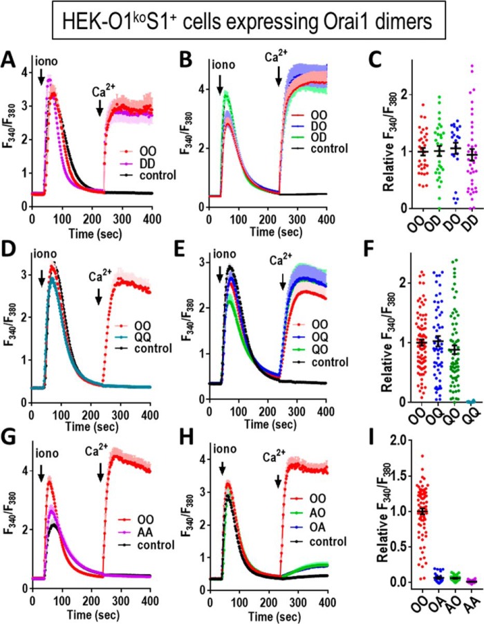
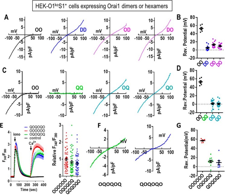
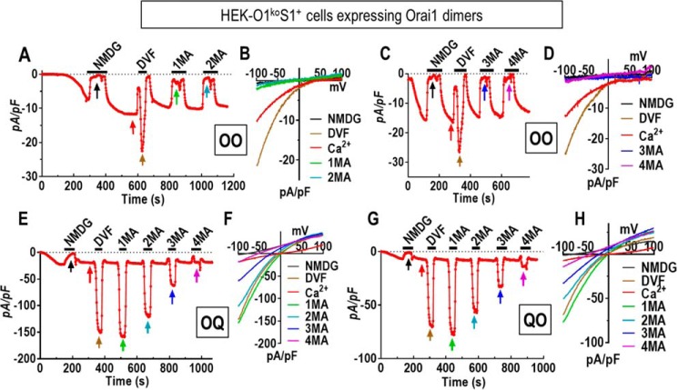
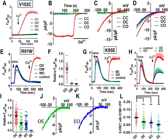
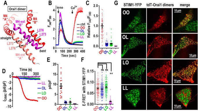
Store-operated Ca2+ entry signals are mediated by plasma membrane Orai channels activated through intermembrane coupling with Ca2+-sensing STIM proteins in the endoplasmic reticulum (ER). The nature of this elaborate Orai-gating mechanism has remained enigmatic. Based on the Drosophila Orai structure, mammalian Orai1 channels are hexamers comprising three dimeric subunit pairs. We utilized concatenated Orai1 dimers to probe the function of key domains within the channel pore and gating regions. The Orai1-E106Q selectivity-filter mutant, widely considered a dominant pore blocker, was surprisingly nondominant within concatenated heterodimers with Orai1-WT. The Orai1-E106Q/WT heterodimer formed STIM1-activated nonselective cation channels with significantly enlarged apparent pore diameter. Other Glu-106 substitutions entirely blocked the function of heterodimers with Orai1-WT. The hydrophobic pore-lining mutation V102C, which constitutively opens channels, was suppressed by Orai1-WT in the heterodimer. In contrast, the naturally occurring R91W pore-lining mutation associated with human immunodeficiency was completely dominant-negative over Orai-WT in heterodimers. Heterodimers containing the inhibitory K85E mutation extending outward from the pore helix gave an interesting partial effect on both channel activation and STIM1 binding, indicating an important allosteric link between the cytosolic Orai1 domains. The Orai1 C-terminal STIM1-binding domain mutation L273D powerfully blocked STIM1-induced channel activation. The Orai1-L273D/WT heterodimer had drastically impaired STIM1-induced channel gating but, unexpectedly, retained full STIM1 binding. This reveals the critical role of Leu-273 in transducing the STIM1-binding signal into the allosteric conformational change that initiates channel gating. Overall, our results provide important new insights into the role of key functional domains that mediate STIM1-induced gating of the Orai1 channel.

Keywords: Orai1; STIM1; calcium; calcium channel; cell signaling; channel gating; ion channel; selectivity filter; store-operated channel.


Conflict of interest statement

The authors declare that they have no conflicts of interest with the contents of this article

Figures

Figure 1.
Figure 1.
Structure of Orai channel and dimeric subunits. A, top view of Drosophila Orai channel hexamer. Two of the six monomers are colored to show one of the three dimers that constitute the channel. The two subunits in each dimer are almost identical except the “A” monomer has a straight M4 extension helix (M4-ext; purple) and the “B” monomer has a bent M4-ext (pink). The two M4-ext helices are linked in an antiparallel configuration by hydrophobic interactions. B, linear sequence of the concatenated Orai1 dimer constructs used; the two monomers are joined through a 36-amino acid linker sequence and tagged with tdTomato at the C terminus. Note: the sequences of the “A” and “B” monomers are the same. C, schematic of the concatenated Orai1 dimer structure. The membrane-spanning domains (M1, M2, M3, M4, and M4-ext) are labeled as shown. Colored dots reveal each of the point mutations used in this study.
Figure 2.
Figure 2.
Consequences of mutating the selectivity filter (Glu-106) within expressed Orai1 monomers. tdTomato-tagged Orai1 monomer constructs were transiently expressed in HEK-O1koS1+ cells that stably express STIM1-YFP. Fura-2 ratiometric Ca2+ measurements were conducted to reveal cytosolic Ca2+ after store depletion with 2.5 μm ionomycin (iono) in Ca2+-free medium followed by addition of 1 mm Ca2+ (arrows). A, cells were transfected with either Orai1-E106D (E106D) or Orai1-WT (O) or untransfected (control). B, cells were co-transfected with both Orai1-E106D-CFP and Orai1 WT or untransfected (control). C, cells were transfected with either Orai1-E106Q (E106Q) or Orai1-WT (O) or untransfected (control). D, cells were co-transfected with both Orai1-E106Q-CFP and Orai1-WT or untransfected (control). All the traces (means ± S.E.) are representative of three independent experiments. Summary scatter plots with means ± S.E. for normalized peak Ca2+ entry are for all individual cells recorded in three independent experiments. (**, p < 0.005).
Figure 3.
Figure 3.
Function of concatenated Orai1 dimers containing the E106D and E106Q selectivity filter mutations. Orai1-tdT dimer constructs were transiently expressed at similar levels in HEK-O1koS1+ cells, and fura-2 ratiometric Ca2+ measurements were made to determine cytosolic Ca2+ after store depletion with 2.5 μm ionomycin (iono) in Ca2+-free medium, followed by addition of 1 mm Ca2+ (arrows). A, cells were transfected with either Orai1-WT homodimer (OO) or Orai1-E106D homodimer (DD) or untransfected (control). B, cells were transfected with either OO or Orai1-WT/Orai1-E106D (OD), Orai1-E106D/Orai1-WT (DO), heterodimers, or untransfected (control). C, summary scatter plots with means ± S.E. for peak Ca2+ entry of all individual cells recorded in three independent experiments with the Orai1 concatemer constructs shown in A and B. D, cells were transfected with either Orai1-WT homodimer (OO) or Orai1-E106Q mutant homodimer (QQ) or were untransfected (control). E, cells were transfected with either OO or with Orai1-WT/Orai1-E106Q (OQ) or Orai1-E106Q/Orai1-WT (QO) dimer or were untransfected (control). F, summary scatter plots with means ± S.E. for peak Ca2+ entry of all individual cells recorded in three independent experiments with the Orai1 concatemer constructs shown in D and E. G, cells were transfected with either Orai1-WT dimer (OO) or Orai1-E106A homodimer (AA) or were untransfected (control). H, cells were transfected with either OO or Orai1-WT/Orai1E106A (OA) or Orai1-E106A/WT dimer (AO) or were untransfected (control). I, summary scatter plots with means ± S.E. for peak Ca2+ entry of all individual cells recorded in three independent experiments with the Orai1 concatemer constructs shown in (G and H). All the traces shown (means ± S.E.) are representative of three independent experiments.
Figure 4.
Figure 4.
Electrophysiological properties of Orai1 E106D and E106Q concatenated dimers. A, representative I/V plots from whole-cell recordings using HEK-O1koS1+ cells transiently expressing each of the tdT-tagged concatenated Orai1 dimer constructs (OO, DD, OD, and DO) as described in Fig. 3. B, summary scatter plot (means ± S.E.) of reversal potential measurements taken from multiple I/V curves for the OO, DD, OD, and DO dimers shown in A. C, representative I/V plots from whole-cell recordings from HEK-O1koS1+ cells transiently expressing each of the tdT-tagged concatenated Orai1 dimer constructs (OO, QQ, OQ, and QO) as described in Fig. 3. D, summary scatter plot (means ± S.E.) of reversal potential measurements taken from multiple I/V curves for the QQ, OQ, and QO as shown in C. Current for the QQ construct was essentially zero. E, fura-2 ratiometric Ca2+ measurements reveal cytosolic Ca2+ after store depletion with 2.5 μm ionomycin (iono) in Ca2+-free medium followed by addition of 1 mm Ca2+ (arrows). In each case, traces for Orai1-WT hexamer (OOOOOO) were compared with traces for Orai1 heterohexamers (OQOQOQ and QOQOQO). Untransfected control cells are also shown. Traces shown (means ± S.E.) are representative of three independent experiments. Summary scatter plots with means ± S.E. for peak Ca2+ entry of all individual cells recorded in three independent experiments with the Orai1 concatemer constructs shown. F, representative I/V relationship for the OQOQOQ and QOQOQO hexamers are shown. G, summary scatter plot (means ± S.E.) of reversal potential measurements taken from multiple I/V curves for the OOOOOO, OQOQOQ, and QOQOQO hexamers as shown in F.
Figure 5.
Figure 5.
Altered pore properties of the nonselective Orai1 concatenated channel dimers OQ and QO. A–D, electrophysiological measurements using HEK-O1koS1+ cells expressing the Orai1-WT dimer (OO). Current developed initially in the presence of 20 mm Ca2+ in the external solution. Thereafter, the external solution was periodically switched from 20 mm Ca2+ with 130 mm Na+ to divalent cation-free solutions containing the following: 150 mm N-methyl-d-glucamine (NMDG), 150 mm Na+ (DVF), 150 mm methylammonium (1MA); 150 mm dimethylammonium (2MA); 150 mm trimethylammonium (3MA); and finally 150 mm tetramethylammonium (4MA). Between each divalent-free addition, cells were switched back to 20 mm Ca2+ with 130 mm Na+. Current at −100 mV was plotted against time, and I/V relationships are shown (B and D) at the times indicated by arrows in A and C. E, using HEK-O1koS1+ cells expressing the OQ dimer, current developed in 20 mm external Ca2+. After switching to NMDG, divalent solutions were again added in the external solution as shown in A and C. For channel formed by OQ heterodimers, methylated ammonium cation still can produce significant currents. F, I/V relationship of currents mediated by OQ collected in different external solutions as indicated by the arrows in E. G, HEK-O1koS1+ cells expressing the QO dimer were exposed to external solutions as shown in E. H, I/V relationship of currents mediated by QO collected in the external solutions indicated in G.
Figure 6.
Figure 6.
Mutation of key Orai1 channel pore residues in concatenated dimers. A, fura-2 ratiometric Ca2+ add-back measurements for HEK-O1ko cells expressing similar levels of the C-terminal tdT-tagged Orai1 dimers OO (Orai1-WT/Orai1-WT), OC (Orai1-WT/Orai1-V102C), (Orai1-V102C/ Orai1-WT), or CC (Orai1-V102C/Orai1-V102C). Constitutive Ca2+ entry was measured after addition of 1 mm Ca2+ (arrows) after Ca2+-free medium. Traces (means ± S.E.) are representative of three independent experiments. B, whole-cell patch–clamp recording of HEK-O1ko cells expressing CC, OC, or CO; current at −100 mV was plotted against time. Cytosolic Ca2+ was maintained at 90 nm with BAPTA to prevent store depletion. Constitutive current was measured in 20 mm Ca2+ external solution, followed by blockade with 10 μm Gd3+. C, representative I/V curves for the constitutive currents of the HEK-O1ko cells expressing OC, CO, and CC shown in B. D, representative I/V curves for Ca2+ currents following store depletion with 20 mm BAPTA in HEK-O1koS1+ cells transiently expressing OC, CO, and CC. E, Ca2+ add-back measurements in fura-2–loaded HEK-O1koS1+ cells expressing similar levels of the C-terminal tdT-tagged Orai1 dimers OO (Orai1-WT/Orai1-WT), OW (Orai1-WT/Orai1-R91W), or WO (Orai1-R91W/Orai1-WT). Ca2+ stores were released with 2.5 μm ionomycin (iono) in Ca2+-free medium followed by 1 mm Ca2+ (arrows). Traces (means ± S.E.) are the results for all cells in three independent experiments. F, summary scatter plots of peak Ca2+ entry normalized to Ca2+ entry with WT dimer (OO); results are means ± S.E. for all cells in three independent experiments represented by E. G and H, Ca2+ add-back measurements using fura-2-loaded HEK-O1koS1+ cells expressing similar levels of the C-terminal tdT-tagged Orai1-K85E homodimers (EE) or the K85E heterodimers (OE and EO). Ca2+ stores were released with 2.5 μm ionomycin in Ca2+-free medium followed by 1 mm Ca2+ (arrows). Traces (means ± S.E.) are representative of all cells in three independent experiments. I, summary scatter plots of peak Ca2+ entry normalized to WT (OO) dimer Ca2+ entry; results are means ± S.E. of three independent experiments represented in G and H. J and K, representative I/V relationship of Ca2+ currents after store depletion in HEK-O1koS1+ cells transiently expressing OE (J) or EO (K). L, scatter plots of E-FRET between stably expressed STIM1-YFP and transiently expressed C-terminally CFP-tagged Orai1 dimers (OO, OE, EO, or EE, respectively) in HEK-O1koS1+ cells. E-FRET was measured at 5 min after addition of 2.5 μm ionomycin to deplete stores. **, p < 0.001. Results are means ± S.E. of three independent experiments.
Figure 7.
Figure 7.
Effects of mutating the key STIM1-binding residue (Leu-273) of the Orai1 C terminus in concatenated Orai1 dimers. A, structural model Orai1 based on the dOrai M4 and M4 extensions (M4-ext), showing their configuration and interactions. The M4-ext from the straight monomer “A” and the M4-ext from the bent monomer “B” lie in a close antiparallel configuration stabilized by interactions between the Leu-273 and Leu-276 residues on each M4-ext. B, Ca2+ entry in fura-2–loaded HEK-O1koS1+ cells expressing similar levels of the C-terminal tdT-tagged Orai1 dimer constructs: OO (Orai1-WT/Orai1-WT), OL (Orai-WT/Orai1-L273D), LO (Orai1-L273D/Orai1-WT), LL (Orai1-L273D/Orai1-L273D), or untransfected control cells (ctrl). Ca2+ stores were released with 2.5 μm ionomycin (iono) in Ca2+-free medium followed by addition of 1 mm Ca2+ (arrows). Traces (means ± S.E.) are representative of three independent experiments. C, summary scatter plots with means ± S.E. of peak Ca2+ entry normalized to peak Ca2+ entry for the OO dimer; all cells are shown from three independent experiments. D, whole-cell patch–clamp recording of HEK-O1koS1+ cells expressing OO, OL, LO, or LL, currents at −100 mV plotted against time. E, summary scatter plots of means ± S.E. of peak Ca2+ current density for all individual cells recorded for each of the Orai1 dimer constructs shown in D. F, scatter plots of E-FRET between stably expressed STIM1-YFP and transiently expressed C-terminally CFP-tagged Orai1 dimers (OO, OL, LO, or LL, respectively) in HEK-O1koS1+ cells. E-FRET was measured at 5 min after addition of 2.5 μm ionomycin to deplete stores. **, p < 0.001; ns, not significant. Results are means ± S.E. of three independent experiments. G, confocal images showing the localization of stably expressed STIM1-YFP and transiently-transfected Orai1-tdTdimers (OO, OL, LO, and LL) expressed in HEK-O1koS1+ cells. Images were taken 5 min after 2.5 μm ionomycin treatment to empty stores. In each case, confocal images show STIM1 and Orai1 dimers localized only in the PM layer immediately adjacent to the coverslip. For OO (top row), there are two cells; the centered cell expresses both STIM1-YFP and OO-tdT, and the lower right cell expresses only STIM1-YFP. Note: there is near-perfect co-localization of STIM and with the OO, OL, and LO Orai1 dimers; in contrast, there is no co-localization of STIM1 with the LL Orai1 dimer.

References

    1. Prakriya M., and Lewis R. S. (2015) Store-operated calcium channels. Physiol. Rev. 95, 1383–1436 10.1152/physrev.00020.2014 - DOI - PMC - PubMed
    1. Amcheslavsky A., Wood M. L., Yeromin A. V., Parker I., Freites J. A., Tobias D. J., and Cahalan M. D. (2015) Molecular biophysics of Orai store-operated Ca channels. Biophys. J. 108, 237–246 10.1016/j.bpj.2014.11.3473 - DOI - PMC - PubMed
    1. Shim A. H., Tirado-Lee L., and Prakriya M. (2015) Structural and functional mechanisms of CRAC channel regulation. J. Mol. Biol. 427, 77–93 10.1016/j.jmb.2014.09.021 - DOI - PMC - PubMed
    1. Zhou Y., Cai X., Nwokonko R. M., Loktionova N. A., Wang Y., and Gill D. L. (2017) The STIM-Orai coupling interface and gating of the Orai1 channel. Cell Calcium 63, 8–13 10.1016/j.ceca.2017.01.001 - DOI - PMC - PubMed
    1. Soboloff J., Rothberg B. S., Madesh M., and Gill D. L. (2012) STIM proteins: dynamic calcium signal transducers. Nat. Rev. Mol. Cell Biol. 13, 549–565 10.1038/nrm3414 - DOI - PMC - PubMed

Publication types

LinkOut - more resources