. 2018 Jun 28;8(1):9573.
doi: 10.1038/s41598-018-27421-1.

Inflammatory cytokines and biofilm production sustain Staphylococcus aureus outgrowth and persistence: a pivotal interplay in the pathogenesis of Atopic Dermatitis

Affiliations

Inflammatory cytokines and biofilm production sustain Staphylococcus aureus outgrowth and persistence: a pivotal interplay in the pathogenesis of Atopic Dermatitis

E G Di Domenico et al. Sci Rep. .

Abstract

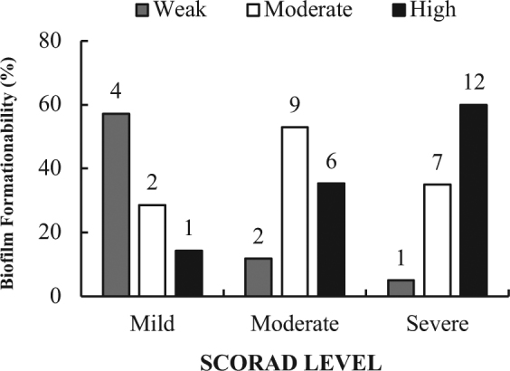
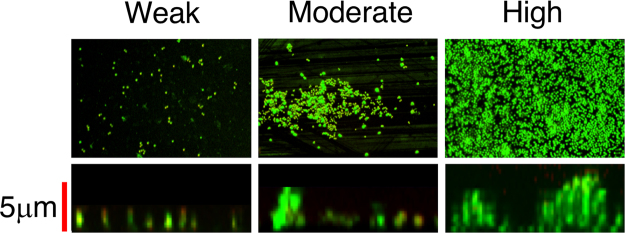
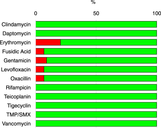
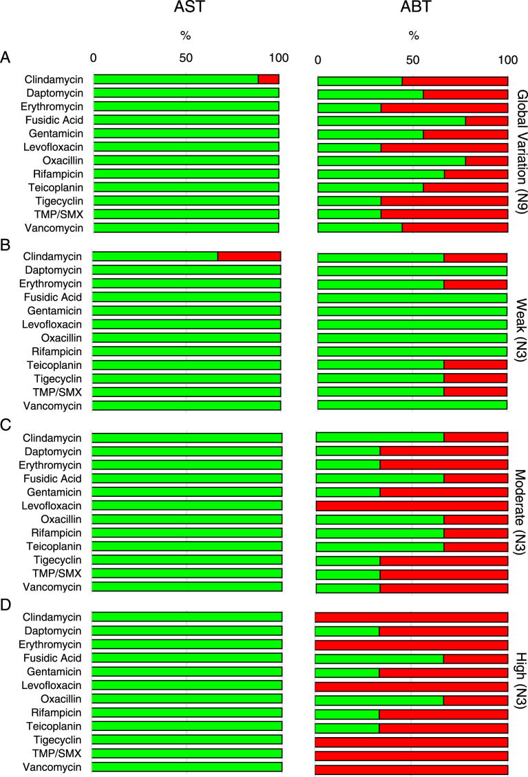
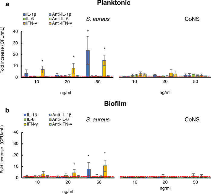
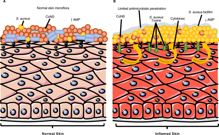
Individuals with Atopic dermatitis (AD) are highly susceptible to Staphylococcus aureus colonization. However, the mechanisms driving this process as well as the impact of S. aureus in AD pathogenesis are still incompletely understood. In this study, we analysed the role of biofilm in sustaining S. aureus chronic persistence and its impact on AD severity. Further we explored whether key inflammatory cytokines overexpressed in AD might provide a selective advantage to S. aureus. Results show that the strength of biofilm production by S. aureus correlated with the severity of the skin lesion, being significantly higher (P < 0.01) in patients with a more severe form of the disease as compared to those individuals with mild AD. Additionally, interleukin (IL)-β and interferon γ (IFN-γ), but not interleukin (IL)-6, induced a concentration-dependent increase of S. aureus growth. This effect was not observed with coagulase-negative staphylococci isolated from the skin of AD patients. These findings indicate that inflammatory cytokines such as IL1-β and IFN-γ, can selectively promote S. aureus outgrowth, thus subverting the composition of the healthy skin microbiome. Moreover, biofilm production by S. aureus plays a relevant role in further supporting chronic colonization and disease severity, while providing an increased tolerance to antimicrobials.


Conflict of interest statement

The authors declare no competing interests.

Figures

Figure 1
Figure 1
Biofilm production by Staphylococcus aureus isolates. Biofilm formation was assessed in the 44S. aureus strains isolated from patients with different degrees of AD. The severity of AD was classified as Mild, Moderate and Severe, respectively, according to the ‘scoring atopic dermatitis’ index (SCORAD). The values above the bars show the the exact number of biofilm-forming isolates. The level of biofilm production was measured by the clinical Biofilm Ring Test (cBRT) and the isolates classified as weak, moderate and high biofilm producers, respectively.
Figure 2
Figure 2
Confocal microscopy images of biofilms. Representative images of biofilms developed on polystyrene pegs following 24 h incubation at 37 °C. Staphylococcus aureus isolates are classified into three groups (weak, moderate and high) according to their biofilm-forming ability. Upper panels show the X-Y planes (top view), while the lower panels show the Z section (side view).
Figure 3
Figure 3
Antimicrobial susceptibility testing for the 44 Staphylococcus aureus isolates. Percentage of susceptible (green) and resistant (red) strains to the indicated antimicrobials, as determined by broth micro-dilution testing. Classification was performed according to the European Committee on Antimicrobial Susceptibility Testing clinical breakpoint tables (EUCAST Clinical Breakpoint Table v 7.1). TXP/SMX - Trimethoprim/Sulfamethoxazole
Figure 4
Figure 4
Comparison between the Antimicrobial susceptibility test (AST) and the Anti-Biofilm Test (ABT). (A) Overall percentage of susceptible (green) and resistant (red) Staphylococcus aureus strains to different antimicrobials as determined in the nine strains (N9). Comparison between AST and ABT in Weak (B), Moderate (C) and High (D) biofilm producing Staphylococcus aureus isolates. Classification was performed according to the European Committee on Antimicrobial Susceptibility Testing clinical breakpoint tables (EUCAST Clinical Breakpoint Table v 7.1). In brackets the number of strains analyzed. TXP/SMX - Trimethoprim/Sulfamethoxazole.
Figure 5
Figure 5
Growth response of Staphylococcus aureus isolates and coagulase-negative staphylococci (CoNS) to different concentrations of cytokines. Planktonic (panel A) and biofilm (panel B) bacteria were cultured overnight in RPMI medium alone (untreated) and in the presence of different concentrations of IL1-β, IL-6 and IFN-γ. Data are expressed as fold increase (CFU/mL) between treated and untreated cells (dashed red line). The strains were tested in triplicate and experiments were repeated at least three times for each cytokine concentration. Mann-Whitney nonparametric test was used for statistical analysis. *Denotes P < 0.05.
Figure 6
Figure 6
Schematic illustration of the pathophysiological changes between normal (A) and inflamed skin in patient with atopic dermatitis (B). Arrows indicate increase (↑) and decrease (↓). The epithelium (https://smart.servier.com/smart_image/epithelium-17/) is adapted from Servier Medical Art (http://smart.servier.com) under the Creative Commons License.

References

    1. Bieber T. Atopic dermatitis. N. Engl. J. Med. 2008;358:1483–1494. doi: 10.1056/NEJMra074081. - DOI - PubMed
    1. Werfel T. The role of leukocytes, keratinocytes, and allergen-specific IgE in the development of atopic dermatitis. J. Invest. Dermatol. 2009;129:1878–1891. doi: 10.1038/jid.2009.71. - DOI - PubMed
    1. Krakowski AC, Eichenfield LF, Dohil MA. Management of atopic dermatitis in the pediatric population. Pediatrics. 2008;122:812–824. doi: 10.1542/peds.2007-2232. - DOI - PubMed
    1. Kezic S, et al. Loss-of-function mutations in the filaggrin gene lead to reduced level of natural moisturizing factor in the stratum corneum. J. Invest. Dermatol. 2008;128:2117–2119. doi: 10.1038/jid.2008.29. - DOI - PubMed
    1. O’Regan GM, et al. Raman profiles of the stratum corneum define 3 filaggrin genotype-determined atopic dermatitis endophenotypes. J. Allergy Clin. Immunol. 2010;126:574–580. doi: 10.1016/j.jaci.2010.04.038. - DOI - PMC - PubMed

MeSH terms