. 2019 Mar;26(3):565-579.
doi: 10.1038/s41418-018-0147-y. Epub 2018 Jun 28.

Enhanced PAPSS2/VCAN sulfation axis is essential for Snail-mediated breast cancer cell migration and metastasis

Affiliations

Enhanced PAPSS2/VCAN sulfation axis is essential for Snail-mediated breast cancer cell migration and metastasis

Yihong Zhang et al. Cell Death Differ. 2019 Mar.

Abstract

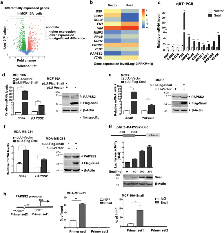
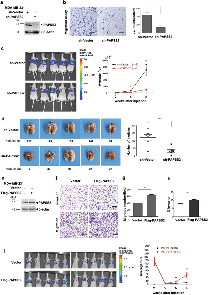
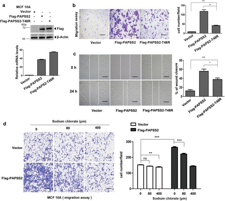
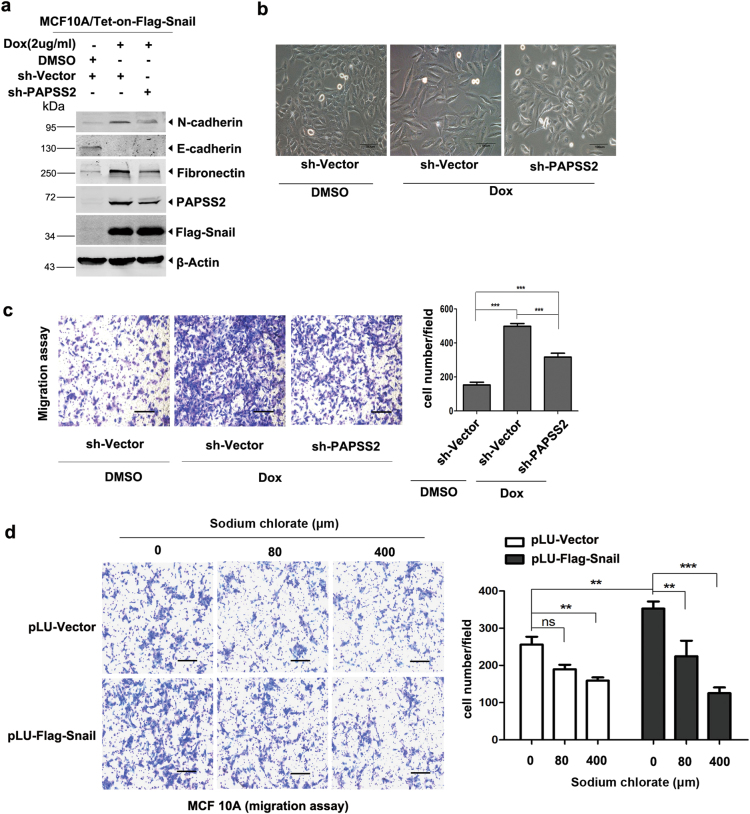
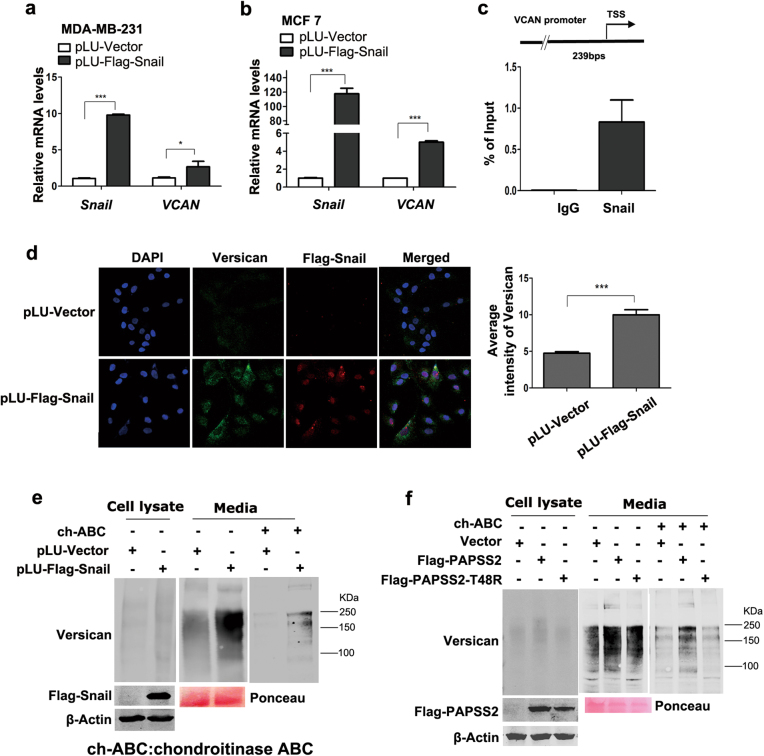
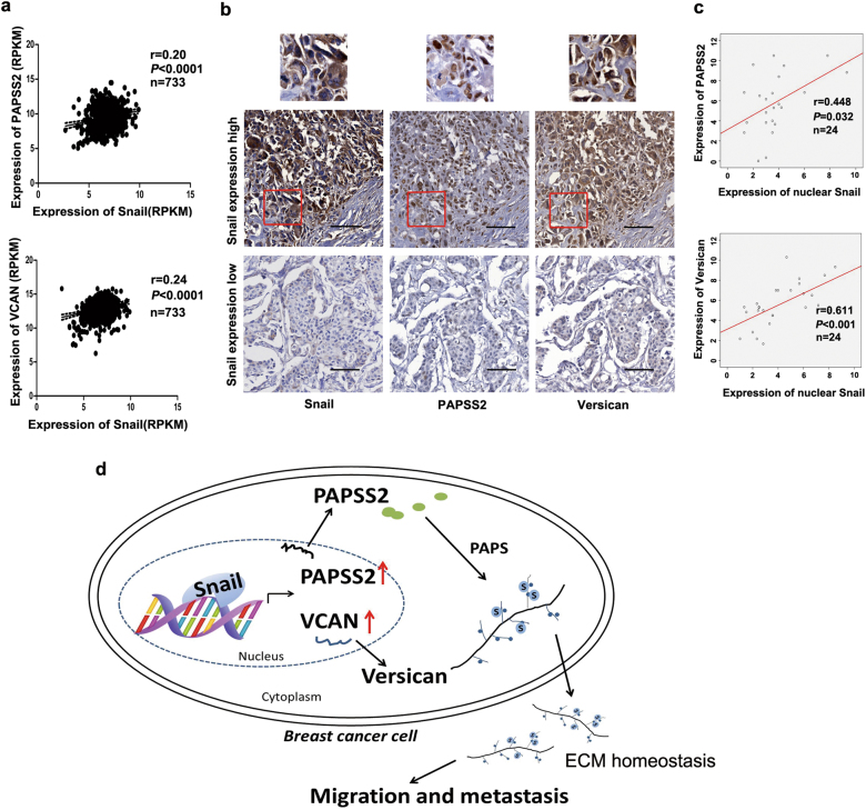
The zinc finger protein Snail is a master regulator of epithelial-mesenchymal transition (EMT) and a strong inducer of tumor metastasis, yet the signal cascades triggered by Snail have not been completely revealed. Here, we report the discovery of the sulfation program that can be induced by Snail in breast cancer cells, and which plays an essential role in cell migration and metastasis. Specifically, Snail induces the expression of PAPSS2, a gene that encodes a rate-limiting enzyme in sulfation pathway, and VCAN, a gene that encodes the chondroitin sulfate proteoglycan Versican in multiple breast cancer cells. Depletion of PAPSS2 in MCF7 and MDA-MB-231 cells results in reduced cell migration, while overexpression of PAPSS2 promotes cell migration. Moreover, MDA-MB-231-shPAPSS2 cells display a significantly lower rate of lung metastasis and lower number of micrometastatic nodules in nude mice, and conversely, MDA-MB-231-PAPSS2 cells increase lung metastasis. Similarly, depletion of VCAN dampens the cell migration activity induced by Snail or PAPSS2 in MCF 10A cells. Moreover, PAPSS inhibitor sodium chlorate effectively decreases cell migration induced by Snail and PAPSS2. More importantly, the expression of Snail, PAPSS2, and VCAN is positively correlated in breast cancer tissues. Together, these findings are important for understanding the genetic programs that control tumor metastasis and may identify previously undetected therapeutic targets to treat metastatic disease.


Conflict of interest statement

The authors declare that they have no conflict of interest.

Figures

Fig. 1
Fig. 1
Snail activates the transcription of PAPSS2 in breast cancer cells. a The Volcano Plot analysis of the differentially expressed genes in MCF 10A-Snail and -vector cells identified by the RNA-seq approach. Red dots represent genes induced by Snail, green dots represent genes repressed by Snail, and blue dots represent genes without significant difference. b Heat map showed the representatives of differentially expressed genes regulated by Snail expression in MCF 10A cells. c qRT-PCR validation of the known Snail target genes in MCF 10A cells. Data were shown as mean ± S.D. from three independent experiments. **P < 0.001, ***P < 0.0001, compared with Vector group. d–f Overexpression of Snail increased PAPSS2 mRNA level in MCF 10A cells (d), MCF7 cells (e), and MDA-MB-231 cells (f). Data were shown as mean ± S.D. from three independent experiments. ***P< 0.0001, **P < 0.001, *P < 0.05, compared with vector group (left panels). Western blots showed the protein levels of Snail and PAPSS2 (right panels). g Human PAPSS2 promoter-luc reporter activity was induced by Snail in 293T cells. Human PAPSS2 gene promoter (−138 +138) was subcloned into pGL3 basic luciferase vector to create a PAPSS2-Luc reporter (upper panel). Assays were performed in 293T cells and the luciferase activity was normalized to β-galactosidase activity. Error bars show standard deviations (bottom panel). h Snail bound to the proximal promoter region of PAPSS2. The ChIP assays were performed in MDA-MB-231 (middle panel) and MCF 10A-Snail cells (right panel) with specific antibody against Snail and the enriched DNA fragments were examined by qPCR. Error bars show standard deviations. **P < 0.001, *P < 0.05
Fig. 2
Fig. 2
PAPSS2 is required for breast cancer cell migration and metastasis. a, e Protein levels of PAPSS2 were quantified by immunoblot analysis in MDA-MB-231 cells with PAPSS2 knocking-down (a) and overexpression (e), respectively. b Transwell assay shows migration abilities of MDA-MB-231-shPAPSS2 cells. Left panel: representative images of migrated cells; right panel: statistical analysis of the average number of migrated cells from the nine randomly chosen fields. Data were shown as mean ± S.D. from three independent experiments. *P < 0.05, compared with Vector groups, 100×, scale bar, 200 μm. c Knocking-down of PAPSS2 in MDA-MB-231 cells inhibited lung metastasis in vivo. Left panel: intensities of lung metastasis in mice at the 6th week were analyzed by in-vivo imaging. Right panel: quantification of lung photon flux at the 2nd, 4th, and 6th week. P value was determined using Student’s t-test, *P < 0.05,**P < 0.001, compared with vector group (n = 7 and n = 6, respectively). d Knocking-down of PAPSS2 decreased number of metastatic nodules on lungs. Left panel: Representative images of metastatic nodules are shown. Right panel: the average number of metastatic tumor nodules shows significant difference between the vector and PAPSS2 knocking-down group. Data were shown as mean ± S.D. ***P < 0.0001, compared with vector group. f–h Migration and invasion capabilities were elevated in PAPSS2-overexpressed MDA-MB-231 cells. f Representative images of invading and migrating cells, 100×, scale bar, 200 μm. g Statistic analysis of migrating cell number, ***P < 0.0001. h Statistic analysis of invasion percentage, % Invasion = (mean number of cell invading through Matrigel Matrix coated membrane/mean number of cells migrating through uncoated membrane) x 100. ***P < 0.0001. i Overexpression of PAPSS2 in MDA-MB-231 cells promoted lung metastasis in vivo. Left panel: intensities of lung metastasis in mice at the 4th week were analyzed by in-vivo imaging. Right panel: quantification of lung photon flux at the 0th, 2nd, 4th and 6th week. P value was determined using Student’s t-test, **P < 0.001, compared with vector group (n = 10)
Fig. 3
Fig. 3
The enzymatic activity is critical for PAPSS2 to induce cell migration. a Upper panel: western blots showed overexpression of PAPSS2 wild type and the mutant T48R in MCF 10A cells. Bottom panel: qRT-PCR analyses showed the mRNA levels of PAPSS2 and T48R. b Left panel: Transwell assays were performed in stable cell lines. Right panel: nine fields chosen randomly were counted for statistical analysis. Data were shown as mean ± S.D. from three independent experiments. **P < 0.001, ***P < 0.0001. c Left panel: Wound healing assay was performed to examine migratory ability, images were taken at 0th, 24th hour post-scratch separately, 40×, scale bar, 500 μm. Right panel: the percentage of wound closure were quantified, wound gaps in six fields chosen randomly were measured at the 0th, 12th, 24th, and 48th hour post-scratch separately. The percentage of wound closure = widths [(0th−24th)/0th * 100%], *P < 0.05, compared with Flag-PAPSS2 group. d Transwell assays showed migration capabilities of PAPSS2-overexpressed cell lines treated with different concentrations of sodium chlorate for 20 h, Right panel: nine fields chosen randomly were counted for statistical analysis. Data were shown as mean ± S.D. from three independent experiments, *P < 0.05, ***P < 0.0001, ns no significance
Fig. 4
Fig. 4
PAPSS2 is an obligate target gene for Snail-induced cell migration. a Western blots showed the protein levels of Snail, PAPSS2, and EMT markers. Depletion of PAPSS2 in Snail-inducible MCF 10A cells. Expression of Snail was induced by Dox (2 μg/ml). b Images showed cellular morphology (200×). Snail induced cell morphological change to mesenchymal type, but knocking-down of PAPSS2 reversed the mesenchymal phenotype into epithelial type in MCF 10A cells. Scale bar, 100 μm. c Transwell assays in MCF 10A-tet-Snail cells with depletion of PAPSS2. Three independent experiments were performed, nine fields chosen randomly were counted for statistical analysis and data were shown as mean ± S.D., ***P < 0.0001. d Transwell assays in Snail-overexpressed MCF 10A cells treated with varied concentration of sodium chlorate for 20 h, **P < 0.001, ***P < 0.0001, ns no significance
Fig. 5
Fig. 5
Snail activates transcription of VCAN and increases sulfation of Versican. a, b Real-time PCR analysis showed increased transcription levels of VCAN in Snail-overexpressed MDA-MB-231 (a) and MCF7 cells lines (b), *P < 0.05, ***P < 0.0001, compared with the vector groups. c Upper: diagram illustrates the human VCAN promoter and the PCR primers used for ChIP. Bottom: examination of the enriched DNA fragments by the ChIP assays using qPCR in MDA-MB-231 cells. Error bars show standard deviations, IgG immunoglobulin. d Snail increased the expression of Versican in MCF 10A cells. Left panel: indirect immunofluorescent assays were performed to examine the subcellular localization of Versican in Snail-overexpressed MCF 10A and MCF 10A-Vector cell lines. Right panel: Average intensity of Versican on every cell in six fields chosen randomly was measured, ***P < 0.0001, compared to the vector group. The average number of cells located in every vision was counted, No = 24/Flag-vector; No = 13/Flag-Snail. e Western blots showed that expression levels of Versican in cell lysate and extracellular matrix, as well as sulfation level in Snail-overexpressed MCF 10A cells. ch-ABC chondroitinase ABC. f Western blots showed that expression levels of Versican in cell lysate and extracellular matrix, as well as sulfation level in PAPSS2-overexpressed MCF 10A cells. Note: Ponceau staining is used as loading control for proteins collected from media
Fig. 6
Fig. 6
Versican is essential for Snail/PAPSS2-induced cell migration. a Western blot showed the protein levels of Versican and Snail when VCAN was knocked down in Snail-overexpressed MCF 10A cells. b Migration capabilities were tested by Transwell assays, depletion of VCAN inhibited Snail-induced cell migration in MCF 10A cells. ***P < 0.0001, compared with vector group. c Western blot showed the protein levels of Versican and PAPSS2 when VCAN was knocked down in PAPSS2-overexpressed MCF 10A cells. d Migration capabilities were tested by Transwell assays, depletion of VCAN inhibited PAPSS2-induced cell migration in MCF 10A cells. ***P < 0.0001, compared with vector group
Fig. 7
Fig. 7
Expression of Snail is positively correlated with the expression of PAPSS2 and Versican in breast cancer specimens. a Scatter plots showed the correlation of Snail expression with PAPSS2 (upper panel) and VCAN (lower panel) expression in 733 breast tumor samples from a TCGA dataset. The r value was calculated via Pearson’s ranking correlation coefficient analysis. b Representative IHC images showed the protein levels of Snail, PAPSS2, and Versican in TMA. Red box indicates cells with amplification. The original magnifications, 200×, scale bar, 100 μm. c Scatter plots showed the correlation of Snail expression with PAPSS2 and Versican expression in TMA samples with positive Snail-nuclear staining. n = 24, the r value was calculated via Pearson’s ranking correlation coefficient analysis. d Schematic model for the role of Snail/PAPSS2/VCAN sulfation axis in breast cancer cell migration and metastasis. Snail activated transcription of PAPSS2 and VCAN via binding to promoter sequence, and simultaneously, induced-PAPSS2 increased sulfation level of Versican, which enhances migration and metastasis of breast cancer cells

Similar articles

Cited by

References

    1. Wang Y, Shi J, Chai K, Ying X, Zhou BP. The role of Snail in EMT and tumorigenesis. Curr Cancer Drug Targets. 2013;13:963–72. doi: 10.2174/15680096113136660102. - DOI - PMC - PubMed
    1. Simpson P. Maternal–zygotic gene interactions during formation of the dorsoventral pattern in Drosophila embryos. Genetics. 1983;105:615–32. - PMC - PubMed
    1. Alberga A, Boulay JL, Kempe E, Dennefeld C, Haenlin M. The snail gene required for mesoderm formation in Drosophila is expressed dynamically in derivatives of all three germ layers. Development. 1991;111:983–92. - PubMed
    1. Moody SE, Perez D, Pan TC, Sarkisian CJ, Portocarrero CP, Sterner CJ, et al. The transcriptional repressor Snail promotes mammary tumor recurrence. Cancer Cell. 2005;8:197–209. doi: 10.1016/j.ccr.2005.07.009. - DOI - PubMed
    1. Brzozowa M, Michalski M, Wyrobiec G, Piecuch A, Dittfeld A, Harabin-Slowinska M, et al. The role of Snail1 transcription factor in colorectal cancer progression and metastasis. Contemp Oncol. 2015;19:265–70. - PMC - PubMed

Publication types

MeSH terms