

Comparison of melanoma gene expression score with histopathology, fluorescence in situ hybridization, and SNP array for the classification of melanocytic neoplasms
- PMID: 29955141
- PMCID: PMC6631355
- DOI: 10.1038/s41379-018-0087-6
Comparison of melanoma gene expression score with histopathology, fluorescence in situ hybridization, and SNP array for the classification of melanocytic neoplasms
Abstract
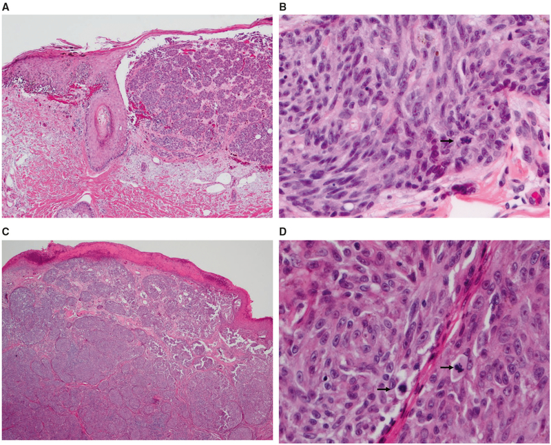
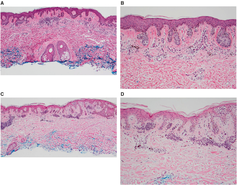
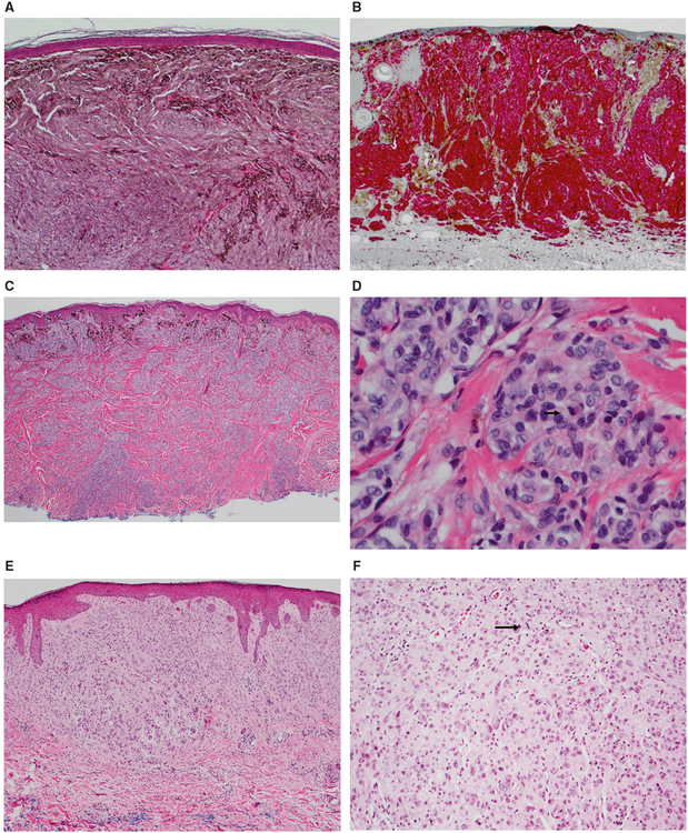
While most melanomas can be distinguished from nevi by histopathology, the histology is ambiguous for some melanocytic tumors, contributing to diagnostic uncertainty. Therefore molecular assays, including FISH or SNP array, and more recently a gene expression test (myPath, Myriad Genetics) have been proposed to aid in the work-up of ambiguous tumors. Two hundred and sixty-eight prospectively submitted cases were gathered, with the goal of comparing the myPath assay to morphologic diagnosis in (1) morphologically unequivocal cases (198), and to morphologic diagnosis and FISH in (2) morphologically ambiguous cases (70). Melanoma FISH was performed using probes for 6p25, 6q23, 11q13, Cep6, 9p21, and Cep9 and scored according to established criteria. The myPath assay was scored by the manufacturer as benign, indeterminate, or malignant. In the unequivocal group, myPath assay showed 75% agreement with morphologic diagnosis, with 67% sensitivity and 81% specificity. In the ambiguous group, FISH and myPath showed 69% inter-test agreement. For these cases agreement with histopathologic interpretation was 84% for FISH and 74% for myPath. Sensitivity and specificity of FISH was 61 and 100%, 50 and 93% for myPath, respectively. Cases from both groups in which myPath was discordant with either morphologic diagnosis and/or FISH (81/268 cases), were submitted for evaluation by two experienced dermatopathologist and also by SNP-array. SNP-array results correlated better than FISH, which correlated better than myPath, with the morphologic interpretation. Our findings document that molecular diagnostics show good correlation with consensus diagnoses, but discordant results occur, and vary in level of correlation with consensus interpretations. Studies with long-term outcomes data within specific ambiguous lesion subsets are required to establish the accuracy of this test, as each molecular diagnostic technique has limitations based on both lack of clinical outcomes data in ambiguous melanocytic tumors and in terms of their sensitivity and specificity in melanocytic lesion subtypes.
Conflict of interest statement
Figures
Comment in
-
Reply to Reimann et al.Mod Pathol. 2019 May;32(5):725-727. doi: 10.1038/s41379-018-0197-1. Epub 2019 Jan 21. Mod Pathol. 2019. PMID: 30666051 Free PMC article. No abstract available.
Similar articles
-
Comparison between melanoma gene expression score and fluorescence in situ hybridization for the classification of melanocytic lesions.Mod Pathol. 2016 Aug;29(8):832-43. doi: 10.1038/modpathol.2016.84. Epub 2016 May 13. Mod Pathol. 2016. PMID: 27174586
-
[Ancillary values of fluorescence in situ hybridization with different gene combination in diagnosis of malignant melanoma].Zhonghua Bing Li Xue Za Zhi. 2020 Aug 8;49(8):827-833. doi: 10.3760/cma.j.issn.cn112151-20200601-00435. Zhonghua Bing Li Xue Za Zhi. 2020. PMID: 32746551 Chinese.
-
Concordance Analysis of the 23-Gene Expression Signature (myPath Melanoma) With Fluorescence In Situ Hybridization Assay and Single Nucleotide Polymorphism Array in the Analysis of Challenging Melanocytic Lesions: Results From an Academic Medical Center.Am J Dermatopathol. 2020 Dec;42(12):939-947. doi: 10.1097/DAD.0000000000001713. Am J Dermatopathol. 2020. PMID: 32675469
-
Fluorescence in situ hybridisation as an ancillary tool in the diagnosis of acral melanoma: a review of 44 cases.Pathology. 2017 Dec;49(7):740-749. doi: 10.1016/j.pathol.2017.08.006. Epub 2017 Oct 14. Pathology. 2017. PMID: 29037804 Review.
-
Nevus versus melanoma: to FISH, or not to FISH.Adv Anat Pathol. 2011 May;18(3):229-34. doi: 10.1097/PAP.0b013e3182169b69. Adv Anat Pathol. 2011. PMID: 21490440 Review.
Cited by
-
Diagnosis of melanoma by imaging mass spectrometry: Development and validation of a melanoma prediction model.J Cutan Pathol. 2021 Dec;48(12):1455-1462. doi: 10.1111/cup.14083. Epub 2021 Jul 2. J Cutan Pathol. 2021. PMID: 34151458 Free PMC article.
-
Molecular Biomarkers for Melanoma Screening, Diagnosis and Prognosis: Current State and Future Prospects.Front Med (Lausanne). 2021 Apr 16;8:642380. doi: 10.3389/fmed.2021.642380. eCollection 2021. Front Med (Lausanne). 2021. PMID: 33937286 Free PMC article. Review.
-
Melanoma pathology: new approaches and classification.Br J Dermatol. 2021 Aug;185(2):282-293. doi: 10.1111/bjd.20427. Epub 2021 May 31. Br J Dermatol. 2021. PMID: 34060071 Free PMC article. Review.
-
Molecular Pathology of Skin Melanoma: Epidemiology, Differential Diagnostics, Prognosis and Therapy Prediction.Int J Mol Sci. 2022 May 11;23(10):5384. doi: 10.3390/ijms23105384. Int J Mol Sci. 2022. PMID: 35628196 Free PMC article. Review.
-
A clinical impact study of dermatologists' use of diagnostic gene expression profile testing to guide patient management.Melanoma Manag. 2024 May 9;11(1):MMT68. doi: 10.2217/mmt-2023-0002. eCollection 2024. Melanoma Manag. 2024. PMID: 38812731 Free PMC article.
References
-
- Cerroni L, Barnhill R, Elder D, et al. Melanocytic tumors of uncertain malignant potential: results of a tutorial held at the XXIX Symposium of the International Society of Dermato-pathology in Graz, October 2008. Am J Surg Pathol. 2010;34:314–26. - PubMed
-
- Scolyer RA, Shaw HM, Thompson JF, et al. Interobserver reproducibility of histopathologic prognostic variables in primary cutaneous melanomas. Am J Surg Pathol. 2003;27:1571–6. - PubMed
-
- Troxel DB. Medicolegal aspects of error in pathology. Arch Pathol Lab Med. 2006;130:617–9. - PubMed
-
- Lang UE, Yeh I, McCalmont TH. Molecular melanoma diagnosis update: gene fusion, genomic hybridization, and massively parallel short-read sequencing. Clin Lab Med. 2017;37:473–84. - PubMed
-
- Curtin JA, Fridlyand J, Kageshita T, et al. Distinct sets of genetic alterations in melanoma. N Engl J Med. 2005;353:2135–47. - PubMed
Publication types
MeSH terms
Grants and funding
LinkOut - more resources
Full Text Sources
Other Literature Sources
Medical
