

Bioactivity-Guided Isolation of Neuritogenic Factor from the Seeds of the Gac Plant (Momordica cochinchinensis)
- PMID: 29955238
- PMCID: PMC6000838
- DOI: 10.1155/2018/8953958
Bioactivity-Guided Isolation of Neuritogenic Factor from the Seeds of the Gac Plant (Momordica cochinchinensis)
Abstract
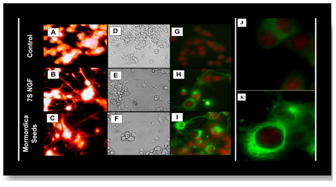
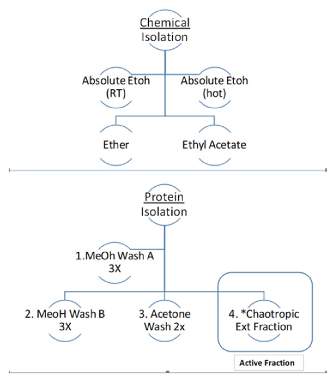
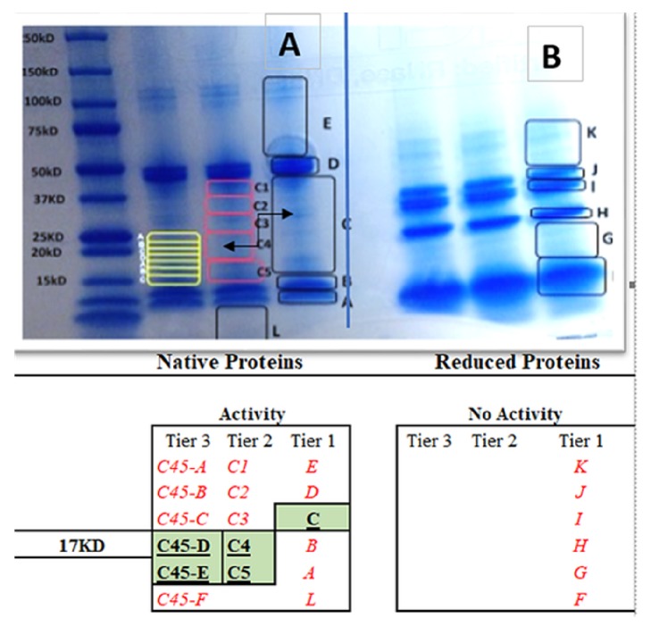
Nerve growth factor (NGF) is an endogenously produced protein with the capacity to induce central nervous system (CNS) neuronal differentiation and repair. NGF signaling involves its binding to tropomyosin-related kinase (Trk) receptors, internalization, and initiation of phosphorylation cascades which cause microtubule reorganization and neuronal outgrowth. Because NGF cannot cross the blood-brain barrier, its therapeutic use is limited. Synthetic peptides that can act as NGF receptor agonists (NGF mimetics) are known to attenuate neurodegenerative pathologies in experimental models of Alzheimer's disease and Parkinson's disease; however, the existence of plant-based NGF mimetics is uncertain. For this reason, we recently completed a high throughput screening of over 1100 nutraceuticals (vitamins, herbal plant parts, polyphenolics, teas, fruits, and vegetables) to identify neuritogenic factor using a PC-12 cell model. Remarkably we found only one, commonly known as the seed of Gac plant (Momordica cochinchinensis) (MCS). In the current study, we further investigated this seed for its neuritogenic effect using bioactivity-guided chemical separations. The data show no biological neuritogenic activity in any chemical solvent fraction, where activity was exclusive to the crude protein. MSC crude proteins were then separated by 1D electrophoresis, where the active neuritogenic activity was confirmed to have a molecular mass of approximately 17 kDa. Subsequently, the 17kDa band was excised, digested, and run on a UPLC-MS/MS with a Q Exactive Hybrid Quadrupole-Orbitrap Mass Spectrometer with data evaluated diverse tools such as X! Tandem, OMS, and K-score algorithms. Proteomic evaluation of the 17kDa band confirmed evidence for 11S globulin subunit beta, napin, oleosin, Momordica trypsin inhibitors (TI) MCoTI-I /II, and many isoforms of Two Inhibitor Peptide Topologies (TIPTOPs). While all peptides identified correspond to the genus/species, Momordica cochinchinensis and Cucumis Sativus, a significant limitation of the analysis is the nonexistence of full annotation for the Momordica cochinchinensis proteome. In conclusion, these findings demonstrate that there is a stable protein within MCS having a mass of 17kDa with the capacity to induce neurite outgrowth. Future work will be required to establish the therapeutic value of the MCS for the treatment of neurodegenerative diseases.
Figures

Similar articles
-
Neurotrophic Effects of Mu Bie Zi (Momordica cochinchinensis) Seed Elucidated by High-Throughput Screening of Natural Products for NGF Mimetic Effects in PC-12 Cells.Neurochem Res. 2015 Oct;40(10):2102-12. doi: 10.1007/s11064-015-1560-y. Epub 2015 Apr 11. Neurochem Res. 2015. PMID: 25862192 Free PMC article.
-
In Silico Investigation of the Binding of MCoTI-II Plant Defense Knottin to the γ-NGF Serine Protease of the 7S Nerve Growth Factor Complex and Biological Activity of Its NGF Mimetic Properties.J Phys Chem B. 2019 Oct 31;123(43):9104-9110. doi: 10.1021/acs.jpcb.9b07547. Epub 2019 Oct 21. J Phys Chem B. 2019. PMID: 31580077 Free PMC article.
-
A comparative study of extraction methods reveals preferred solvents for cystine knot peptide isolation from Momordica cochinchinensis seeds.Fitoterapia. 2014 Jun;95:22-33. doi: 10.1016/j.fitote.2014.02.016. Epub 2014 Mar 5. Fitoterapia. 2014. PMID: 24613804
-
Phytochemistry, Pharmacological Activities, Toxicity and Clinical Application of Momordica cochinchinensis.Curr Pharm Des. 2019;25(6):715-728. doi: 10.2174/1381612825666190329123436. Curr Pharm Des. 2019. PMID: 30931848 Review.
-
Behind the Myth of the Fruit of Heaven, a Critical Review on Gac (Momordica cochinchinensis Spreng.) Contribution to Nutrition.Curr Med Chem. 2019;26(24):4585-4605. doi: 10.2174/0929867326666190705154723. Curr Med Chem. 2019. PMID: 31284852 Review.
Cited by
-
In Silico, Molecular Docking and In Vitro Antimicrobial Activity of the Major Rapeseed Seed Storage Proteins.Front Pharmacol. 2020 Sep 8;11:1340. doi: 10.3389/fphar.2020.01340. eCollection 2020. Front Pharmacol. 2020. PMID: 33013372 Free PMC article.
-
Selected Seeds as Sources of Bioactive Compounds with Diverse Biological Activities.Nutrients. 2022 Dec 30;15(1):187. doi: 10.3390/nu15010187. Nutrients. 2022. PMID: 36615843 Free PMC article. Review.
-
A Potential Anti-Tumor Herb Bred in a Tropical Fruit: Insight into the Chemical Components and Pharmacological Effects of Momordicae Semen.Molecules. 2019 Oct 31;24(21):3949. doi: 10.3390/molecules24213949. Molecules. 2019. PMID: 31683690 Free PMC article. Review.
References
Grants and funding
LinkOut - more resources
Full Text Sources
Other Literature Sources
