Case Reports
. 2018 May 31:2018:6815407.
doi: 10.1155/2018/6815407. eCollection 2018.

Neurotrophic Keratopathy after Trigeminal Nerve Block for Treatment of Postherpetic Neuralgia

Affiliations
Case Reports

Neurotrophic Keratopathy after Trigeminal Nerve Block for Treatment of Postherpetic Neuralgia

Aya Kodama-Takahashi et al. Case Rep Ophthalmol Med. .

Abstract

Purpose: To report a case of persistent corneal epithelial defect that had occurred after a trigeminal nerve block.

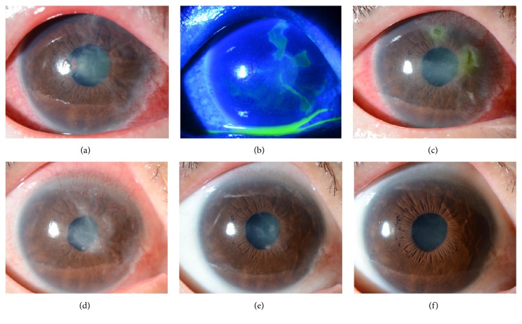
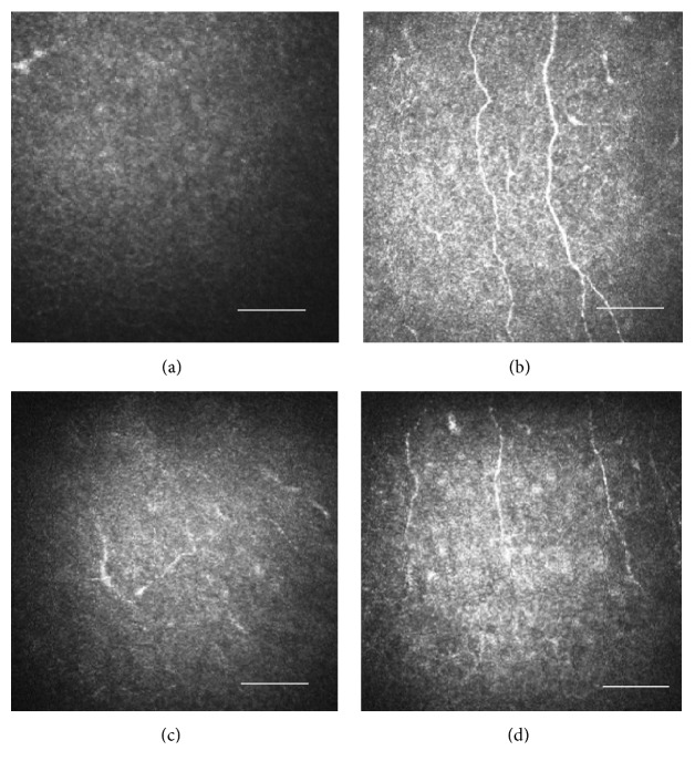
Case presentation: A 75-year-old female had suffered from postherpetic neuralgia for 8 years. She underwent Gasserian ganglion block surgery and noticed declining visual acuity in the right eye on the following day. She presented with severe hyperemia and corneal epithelial defects in the right eye and experienced remarkable reduction of sensitivity in the right cornea. She was diagnosed with neurotrophic keratopathy. Ofloxacin eye ointment and rebamipide ophthalmic suspension ameliorated the corneal epithelial defects but superficial punctate keratopathy, corneal superficial neovascularization, and Descemet's fold persisted. Although the epithelial defects occasionally recurred, the corneal sensation and epithelial defects, Descemet's fold, and corneal superficial neovascularization all improved around 5 months after trigeminal nerve block. The HRT II Rostock Cornea Module (RCM) could not detect any corneal subbasal nerve fibers at postoperative 4 months; however, it could detect them at postoperative 6 months.

Conclusions: As the nerve block effect wore off, the corneal subbasal nerve fibers slowly regenerated. As the corneal sensation improved, the corneal epithelial defects and superficial neovascularization also improved. The HRT II RCM appeared useful for observing loss and regeneration of the corneal subbasal nerve fibers.


Figures

Figure 1
Figure 1
Slit-lamp images of the right eye showing the clinical course.(a) One week after the trigeminal nerve block, corneal epithelial defects, severe conjunctival hyperemia all around the periphery, cloudy and irregular corneal epithelium and Descemet's membrane folds, and mild stromal edema were noted. (b) One week after the trigeminal nerve block, the corneal epithelial defects were shown using fluorescein staining. (c) One month after the nerve block, PED were noted. Severe conjunctival hyperemia, very severe corneal superficial neovascularization at 11 to 5 o'clock, and corneal stromal edema in the central cornea were also observed. (d) Two months after the trigeminal nerve block, the epithelial defects were improved but SPK, corneal superficial neovascularization at 11 to 5 o'clock, irregularity of corneal epithelium, and stromal scarring in the central cornea remained. (e) Five months after the nerve block, slight irregularity of corneal epithelium and stromal scarring in the central cornea were observed. Corneal superficial neovascularization showed signs of improvement although SPK remained. (f) Six months after the nerve block, SPK and conjunctival hyperemia, corneal superficial neovascularization, and corneal stromal edema were all improved.
Figure 2
Figure 2
Evaluation of the subbasal nerve fibers in the cornea with the HRT II RCM. Size bar = 100 μm. (a) Four months after the nerve block, the subbasal nerve fibers were hardly observed in the right eye. (b) Four months after the nerve block, the subbasal nerve fibers were observed in the fellow eye. (c) Six months after the nerve block, regenerated subbasal nerve fibers were observed starting from the periphery of the right cornea, although they appeared small and short. (d) Six months after the nerve block, subbasal nerve fibers were observed in the fellow eye.

References

    1. Nishida T. Neurotrophic mediators and corneal wound healing. The Ocular Surface. 2005;3(4):194–202. doi: 10.1016/S1542-0124(12)70206-9. - DOI - PubMed
    1. Nishida T., Inui M., Nomizu M. Peptide therapies for ocular surface disturbances based on fibronectin-integrin interactions. Progress in Retinal and Eye Research. 2015;47:38–63. doi: 10.1016/j.preteyeres.2015.01.004. - DOI - PubMed
    1. Okada Y., Reinach P. S., Kitano A., et al. Neurotrophic keratopathy; its pathophysiology and treatment. Histol Histopathol. 2010;25(6):771–80. - PubMed
    1. Sacchetti M., Lambiase A. Diagnosis and management of neurotrophic keratitis. Clinical Ophthalmology. 2014;19(8):571–579. doi: 10.2147/opth.s45921. - DOI - PMC - PubMed
    1. Lambiase A., Sacchetti M., Mastropasqua A., Bonini S. Corneal changes in neurosurgically induced neurotrophic keratitis. JAMA Ophthalmology. 2013;131(12):1547–1553. doi: 10.1001/jamaophthalmol.2013.5064. - DOI - PubMed

Publication types

LinkOut - more resources