. 2018 May 14;7(6):e623-e632.
doi: 10.1016/j.eats.2018.02.009. eCollection 2018 Jun.

Arthroscopic Bone Graft Procedure Combined With Arthroscopic Subscapularis Augmentation for Recurrent Anterior Instability With Glenoid Bone Defect

Affiliations

Arthroscopic Bone Graft Procedure Combined With Arthroscopic Subscapularis Augmentation for Recurrent Anterior Instability With Glenoid Bone Defect

Raffaele Russo et al. Arthrosc Tech. .

Abstract

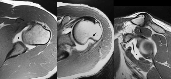
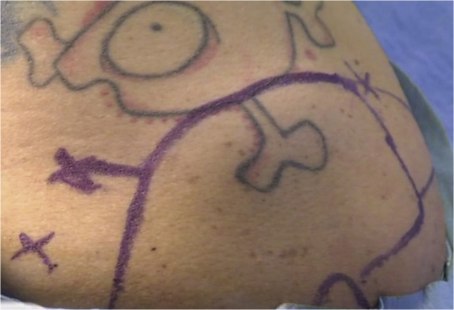
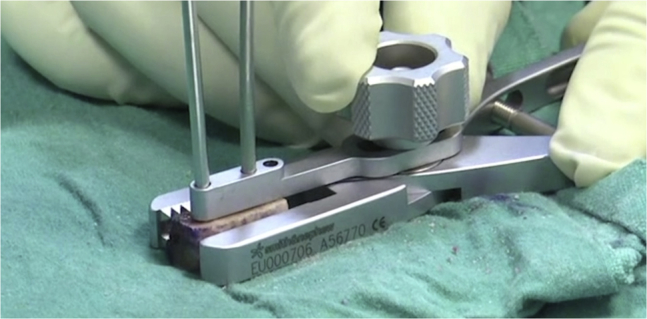
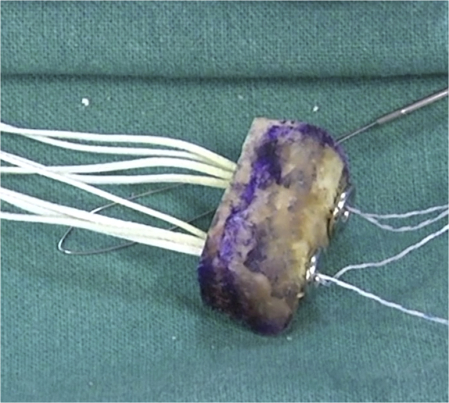
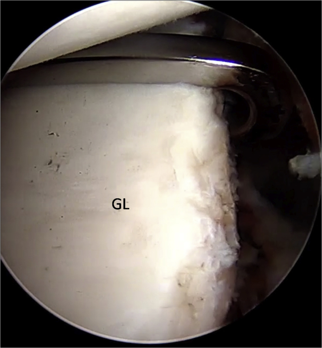
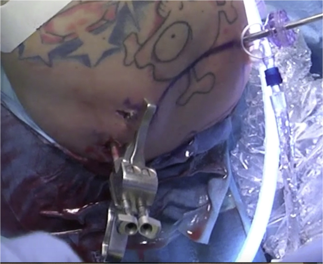
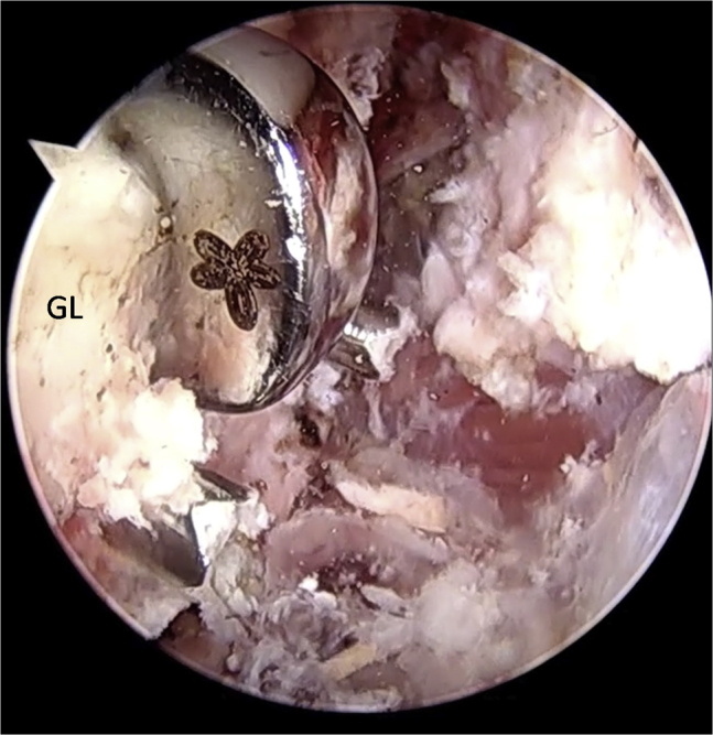
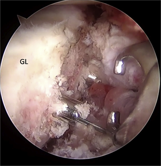
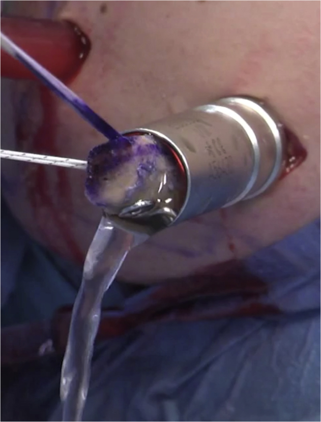
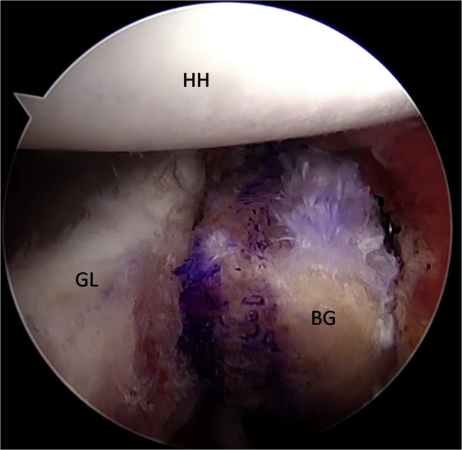
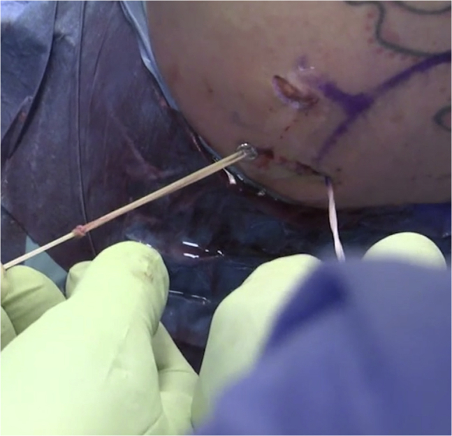
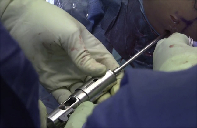
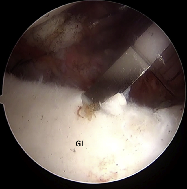
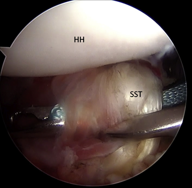
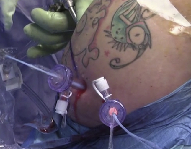
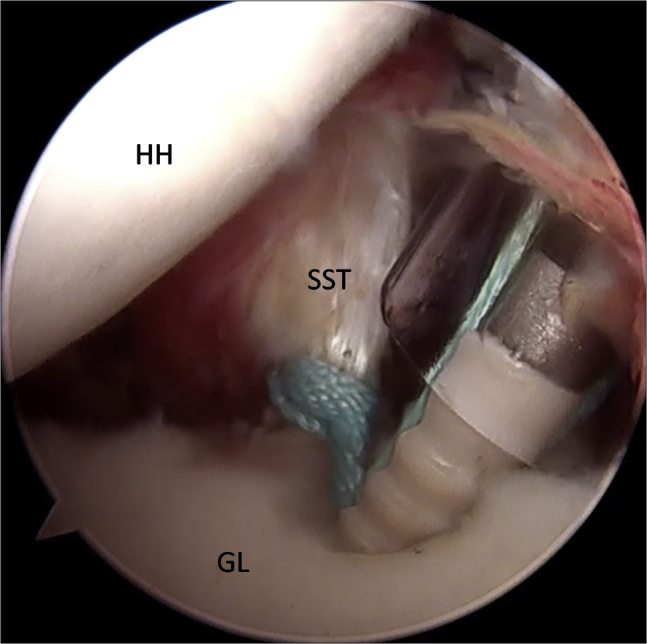
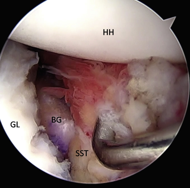
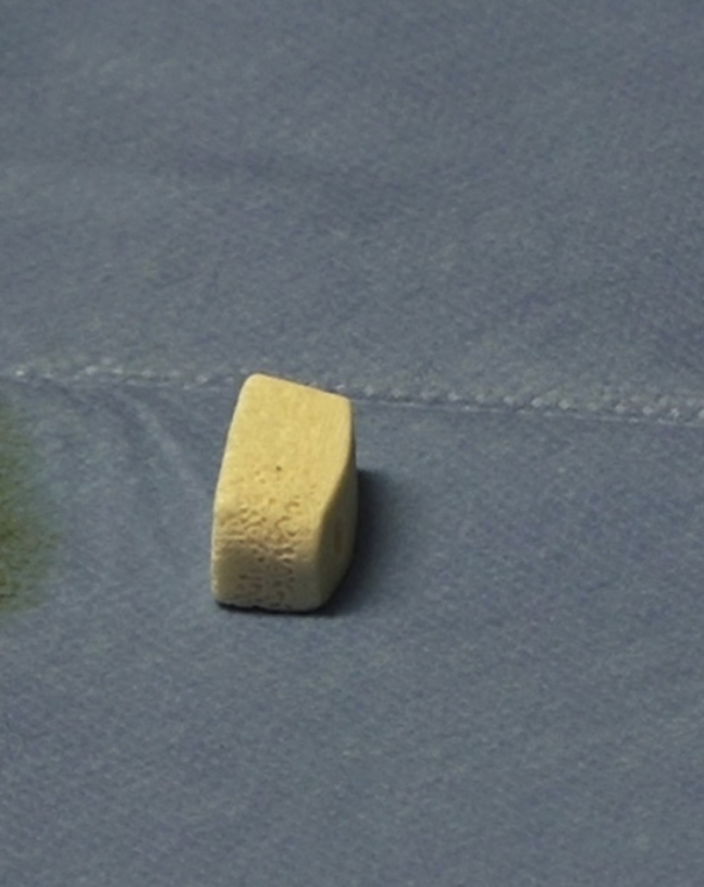
Glenoid bone loss and capsular deficiency represent critical points of arthroscopic Bankart repair failures. The purpose of this Technical Note is to present an all-arthroscopic bone block procedure associated with arthroscopic subscapularis augmentation for treating glenohumeral instability with glenoid bone loss and anterior capsulolabral deficiency. Two glenoid tunnels are set up from the posterior to the anterior side using a dedicated bone block guide, and 4 buttons are used to fix the graft to the glenoid. The subscapularis tenodesis is performed using a suture tape anchor. This combined arthroscopic technique (bone block associated with arthroscopic subscapularis augmentation) could be a valid and safe alternative to the arthroscopic or open Latarjet procedures.


Figures

Fig 1
Fig 1
Arthro-MRI is useful to detect Hill-Sachs lesions (arrow) and anterior capsulolabral deficiency (asterisk). Left shoulder, axial/axial/sagittal view. (MRI, magnetic resonance imaging.)
Fig 2
Fig 2
A posterior portal is created 1 cm lateral to the standard portal for the introduction of the glenoid drilling guide.
Fig 3
Fig 3
Two 2.8-mm drill holes are made 10 mm apart and 5 mm from each edge of the graft.
Fig 4
Fig 4
The graft preassembled with the suture.
Fig 5
Fig 5
Left shoulder, anterior on the right and posterior on the left, an arthroscope from the anterosuperior portal, and a hook guide from the posterior portal. The hook of the guide is passed parallel to the glenoid face, and it is advanced over the anterior edge. (GL, glenoid.)
Fig 6
Fig 6
Left shoulder, view from posterior. Once the guide is positioned and aligned with the posterior and anterior glenoid rims, a bullet is placed in each hole of the guide.
Fig 7
Fig 7
Left shoulder, anterior on the right and posterior on the left, an arthroscope from the anterosuperior portal. The inner drill is removed leaving the cannulated outer sleeve. (GL, glenoid.)
Fig 8
Fig 8
Left shoulder, anterior on the right and posterior on the left, an arthroscope from the anterosuperior portal, and a loop grasper from the anteroinferior portal. Each flexible looped guidewire, introduced into the joint by passing 1 wire through each sleeve in a posterior to anterior direction, is retrieved using a loop grasper. (GL, glenoid.)
Fig 9
Fig 9
Left shoulder, view from anterior, a metal cannula in the anteroinferior portal, and an arthroscopic cannula in the anterosuperior portal. The graft with the preassembled suture is advanced through a 15-mm metal cannula.
Fig 10
Fig 10
Left shoulder, anterior on the right and posterior on the left, an arthroscope from the anterosuperior portal. The graft is advanced until it sits flush on the glenoid neck. (BG, bone graft; GL, glenoid; HH, humeral head.)
Fig 11
Fig 11
Left shoulder, view from posterior. The posterior round endobuttons are advanced using a sliding knot until they are flush with the posterior face of the glenoid.
Fig 12
Fig 12
Left shoulder, view from posterior. The suture tensioner device is used to secure the posterior round endobuttons.
Fig 13
Fig 13
Left shoulder, anterior on the right and posterior on the left, an arthroscope from the anterosuperior portal. A suture anchor bone hole is made at 10 o'clock position on the anterior glenoid edge. (GL, glenoid.)
Fig 14
Fig 14
Left shoulder, anterior on the right and posterior on the left, an arthroscope from the posterior portal, and a suture-passing device from the anteroinferior portal. The middle upper third of the subscapularis tendon is penetrated with a suture-passing device loaded with the tape. (HH, humeral head; SST, subscapularis tendon.)
Fig 15
Fig 15
Left shoulder, view from anterior. The tape is carried out through the cannula with a suture retriever and then passed again in the other cannula.
Fig 16
Fig 16
Left shoulder, view from anterior. Both free ends of the tape are passed through the 2.9-mm anchor's eyelet and the anchor is pushed along the tape toward the bone hole from the anteroinferior portal.
Fig 17
Fig 17
Left shoulder, anterior on the right and posterior on the left, an arthroscope from the posterior portal, and an anchor from the anteroinferior portal. The anchor is inserted into the bone. (GL, glenoid; HH, humeral head; SST, subscapularis tendon.)
Fig 18
Fig 18
Left shoulder. Final view from the anterosuperior portal showing the closure of the anterior pounce on the bone graft and subscapularis tenodesis. (BG, bone graft; GL, glenoid; HH, humeral head; SST, subscapularis tendon.)
Fig 19
Fig 19
The preformed Xenograft.

References

    1. Alkaduhimi H., van der Linde J.A., Willigenburg N.W., Paulino Pereira N.R., van Deurzen D.F., van den Bekerom M.P. Redislocation risk after an arthroscopic Bankart procedure in collision athletes: A systematic review. J Shoulder Elbow Surg. 2016;25:1549–1558. - PubMed
    1. Arciero R.A., Parrino A., Bernhardson A.S. The effect of a combined glenoid and Hill-Sachs defect on glenohumeral stability: A biomechanical cadaveric study using 3-dimensional modeling of 142 patients. Am J Sports Med. 2015;43:1422–1429. - PubMed
    1. Burkhart S.S., De Beer J.F. Traumatic glenohumeral bone defects and their relationship to failure of arthroscopic Bankart repairs: Significance of the inverted-pear glenoid and the humeral engaging Hill-Sachs lesion. Arthroscopy. 2000;16:677–694. - PubMed
    1. Di Giacomo G., de Gasperis N., Scarso P. Bipolar bone defect in the shoulder anterior dislocation. Knee Surg Sports Traumatol Arthrosc. 2016;24:479–488. - PubMed
    1. Shin S.J., Koh Y.W., Bui C. What is the critical value of glenoid bone loss at which soft tissue Bankart repair does not restore glenohumeral translation, restricts range of motion, and leads to abnormal humeral head position? Am J Sports Med. 2016;44:2784–2791. - PubMed

LinkOut - more resources