Annatto Tocotrienol Attenuates NLRP3 Inflammasome Activation in Macrophages
- PMID: 29955706
- PMCID: PMC5998354
- DOI: 10.3945/cdn.117.000760
Annatto Tocotrienol Attenuates NLRP3 Inflammasome Activation in Macrophages
Abstract
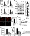
Accumulating evidence suggests that aberrant innate immunity is closely linked to metabolic diseases, including type 2 diabetes. In particular, activation of the NOD-like receptor family pyrin domain-containing 3 (NLRP3) inflammasome and subsequent secretion of interleukin 1β (IL-1β) are critical determinants that precipitate disease progression. The seeds of annatto (Bixa orellana L.) contain tocotrienols (T3s), mostly (>90%) in the δ form (δT3). The aim of this study was to determine whether annatto T3 is effective in attenuating NLRP3 inflammasome activation in macrophages. Our results showed that annatto δT3 significantly attenuated NLRP3 inflammasome by decreasing IL-1β reporter activity, IL-1β secretion, and caspase-1 cleavage against lipopolysaccharide (LPS) followed by nigericin stimulation. With regard to mechanism, annatto δT3 1) reduced LPS-mediated priming of the inflammasome and 2) dampened reactive oxygen species production, the second signal required for assembly of the NLRP3 inflammasome in macrophages. Our work suggests that annatto δT3 may hold therapeutic potential for delaying the onset of NLRP3 inflammasome-associated chronic metabolic diseases.
Keywords: IL-1β; NLRP3 inflammasome; ROS production; annatto; delta-tocotrienol.
Figures


Similar articles
-
[Inhibitory effect and mechanism of deoxyschizandrin on NLRP3 inflammasome].Yao Xue Xue Bao. 2017 Jan;52(1):80-5. Yao Xue Xue Bao. 2017. PMID: 29911779 Chinese.
-
Forskolin attenuates the NLRP3 inflammasome activation and IL-1β secretion in human macrophages.Pediatr Res. 2019 Dec;86(6):692-698. doi: 10.1038/s41390-019-0418-4. Epub 2019 May 13. Pediatr Res. 2019. PMID: 31086288
-
Koumine Suppresses IL-1β Secretion and Attenuates Inflammation Associated With Blocking ROS/NF-κB/NLRP3 Axis in Macrophages.Front Pharmacol. 2021 Jan 18;11:622074. doi: 10.3389/fphar.2020.622074. eCollection 2020. Front Pharmacol. 2021. PMID: 33542692 Free PMC article.
-
Genetic and Epigenetic Regulation of the Innate Immune Response to Gout.Immunol Invest. 2023 Apr;52(3):364-397. doi: 10.1080/08820139.2023.2168554. Epub 2023 Feb 6. Immunol Invest. 2023. PMID: 36745138 Review.
-
Annatto (Bixa orellana)-Based Nanostructures for Biomedical Applications-A Systematic Review.Pharmaceutics. 2024 Sep 29;16(10):1275. doi: 10.3390/pharmaceutics16101275. Pharmaceutics. 2024. PMID: 39458606 Free PMC article. Review.
Cited by
-
Vitamin E δ-tocotrienol inhibits TNF-α-stimulated NF-κB activation by up-regulation of anti-inflammatory A20 via modulation of sphingolipid including elevation of intracellular dihydroceramides.J Nutr Biochem. 2019 Feb;64:101-109. doi: 10.1016/j.jnutbio.2018.10.013. Epub 2018 Nov 3. J Nutr Biochem. 2019. PMID: 30471562 Free PMC article.
-
The Vitamin E Derivative Garcinoic Acid Suppresses NLRP3 Inflammasome Activation and Pyroptosis in Murine Macrophages.Inflammation. 2025 Feb 21. doi: 10.1007/s10753-025-02269-6. Online ahead of print. Inflammation. 2025. PMID: 39982672
-
Revisiting the therapeutic potential of tocotrienol.Biofactors. 2022 Jul;48(4):813-856. doi: 10.1002/biof.1873. Epub 2022 Jun 20. Biofactors. 2022. PMID: 35719120 Free PMC article. Review.
-
The Effects of Annatto Tocotrienol Supplementation on Cartilage and Subchondral Bone in an Animal Model of Osteoarthritis Induced by Monosodium Iodoacetate.Int J Environ Res Public Health. 2019 Aug 13;16(16):2897. doi: 10.3390/ijerph16162897. Int J Environ Res Public Health. 2019. PMID: 31412648 Free PMC article.
-
Tocotrienols: Dietary Supplements for Chronic Obstructive Pulmonary Disease.Antioxidants (Basel). 2021 May 31;10(6):883. doi: 10.3390/antiox10060883. Antioxidants (Basel). 2021. PMID: 34072997 Free PMC article. Review.
References
-
- Hernandez J-C, Sirois CM, Latz E.. Activation and regulation of the NLRP3 inflammasome. Couillin I, Pétrilli V, Martinon F. editors. The inflammasomes. Basel (Switzerland): Springer Basel; 2011. p. 197–208.
-
- Tennant DR, O'Callaghan M.. Survey of usage and estimated intakes of annatto extracts. Food Res Int 2005;38:911–7.
LinkOut - more resources
Full Text Sources
Other Literature Sources

