. 2018 Sep;16(9):1873-1886.
doi: 10.1111/jth.14218. Epub 2018 Aug 12.

Tuning the endothelial response: differential release of exocytic cargos from Weibel-Palade bodies

Affiliations

Tuning the endothelial response: differential release of exocytic cargos from Weibel-Palade bodies

T D Nightingale et al. J Thromb Haemost. 2018 Sep.

Abstract

Essentials Endothelial activation initiates multiple processes, including hemostasis and inflammation. The molecules that contribute to these processes are co-stored in secretory granules. How can the cells control release of granule content to allow differentiated responses? Selected agonists recruit an exocytosis-linked actin ring to boost release of a subset of cargo.

Summary: Background Endothelial cells harbor specialized storage organelles, Weibel-Palade bodies (WPBs). Exocytosis of WPB content into the vascular lumen initiates primary hemostasis, mediated by von Willebrand factor (VWF), and inflammation, mediated by several proteins including P-selectin. During full fusion, secretion of this large hemostatic protein and smaller pro-inflammatory proteins are thought to be inextricably linked. Objective To determine if secretagogue-dependent differential release of WPB cargo occurs, and whether this is mediated by the formation of an actomyosin ring during exocytosis. Methods We used VWF string analysis, leukocyte rolling assays, ELISA, spinning disk confocal microscopy, high-throughput confocal microscopy and inhibitor and siRNA treatments to demonstrate the existence of cellular machinery that allows differential release of WPB cargo proteins. Results Inhibition of the actomyosin ring differentially effects two processes regulated by WPB exocytosis; it perturbs VWF string formation but has no effect on leukocyte rolling. The efficiency of ring recruitment correlates with VWF release; the ratio of release of VWF to small cargoes decreases when ring recruitment is inhibited. The recruitment of the actin ring is time dependent (fusion events occurring directly after stimulation are less likely to initiate hemostasis than later events) and is activated by protein kinase C (PKC) isoforms. Conclusions Secretagogues differentially recruit the actomyosin ring, thus demonstrating one mechanism by which the prothrombotic effect of endothelial activation can be modulated. This potentially limits thrombosis whilst permitting a normal inflammatory response. These results have implications for the assessment of WPB fusion, cargo-content release and the treatment of patients with von Willebrand disease.

Keywords: Weibel-Palade bodies; exocytosis; hemostasis; inflammation; von Willebrand factor.


Figures

Figure 1
Figure 1
The actomyosin ring increases the efficiency of von Willebrand factor (VWF) string formation but has little effect on leukocyte rolling. Human umbilical vein endothelial cells (HUVECs) stimulated under flow with histamine (100 μmol L−1)/adrenalin (10 μmol L−1)/IBMX (100 μmol L−1) in the presence or absence of 0.25 μmol L−1 cytochalasin E (CCE) and fixed for string length analysis (A–C) or perfused with THP‐1 leukocytes for rolling analysis (D) (n = 3). (A, B) HUVECs were fixed and stained for VWF before imaging on a confocal microscope; pictures shown are tile scans of 10 fields of view. The whole image (i) and with the boxed area magnified (ii) are shown with a filter added to improve contrast. Scale bar 50 μm. (C) The lengths of VWF strings were quantified from three independent experiments (control, n = 13 images, 1346 strings; CCE, n = 14 images, 1364 strings). The percentage of strings less than 25 μm, between 25 and 50 μm and longer than 50 μm was calculated per image and standard error of the mean (SEM) shown. (D) The number of interacting THP‐1 leukocytes/min was determined from movies. Each point represents the total number of interacting leukocytes per 1‐min movie, with up to two movies acquired per experiment from stimulated cells. Error bars represent standard deviation (SD). Statistical significance assessed using the Mann–Whitney test (C) and one‐way anova with Dunnet's multiple comparison test (D). *P ≤ 0.05.
Figure 2
Figure 2
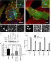
Different secretagogues release von Willebrand factor (VWF) and VWF pro‐peptide with differing efficiencies in a manner that is dependent on the actomyosin ring. (A) Human umbilical vein endothelial cells (HUVECs) were stimulated with 100 ng mL−1 phorbol 12‐myristate 13‐acetate (PMA) for 5 min and fixed using a procedure optimal for the actin cytoskeleton, co‐stained for VWF (red) and phalloidin (green) and imaged on a confocal microscope. Maximum intensity projections shown. Boxed regions are shown magnified. Bar 10 μm. (B) Schematic of Weibel‐Palade body exocytosis in the presence or absence of an actomyosin ring. Small cargo release is ring independent, whereas VWF release is more efficient in the presence of the ring. (C) Quantification of PMA (100 ng mL−1), histamine (100 μmol L−1) or histamine (100 μmol L−1)/adrenalin (10 μmol L−1)/3‐isobutyl‐1‐methyl xanthine (100 μmol L−1)‐stimulated (Ci) VWF or (Cii) pro‐peptide secretion (n = 6–9); error bars = SEM. (Ciii) Ratio of stimulated VWF:propeptide release. Boxes represent 25th–75th percentiles; whiskers represent minimum and maximum values. (D) Quantification of PMA (100 ng mL−1)‐stimulated (Di) VWF or (Dii) pro‐peptide secretion in the presence or absence of 25 μmol L−1 blebbistatin or 1 μmol L−1 cytochalasin E (n = 4); error bars = standard error of the mean (SEM). (Diii) Ratio of stimulated VWF:propeptide release. Error bars = SEM. Statistical significance assessed using t‐test with Welch's correction (Ci‐ii and Di‐ii) and ratio t‐test (Ciii and Diii). *P ≤ 0.05, **P ≤ 0.01 and ***P ≤ 0.001. [Color figure can be viewed at http://wileyonlinelibrary.com]
Figure 3
Figure 3
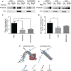
Release of P‐selectin from WPBs for recruitment to the plasma membrane is partially ring dependent. The proportion of cell surface to total P‐selectin levels was determined by surface biotinylation and neutravidin pulldown following stimulation with PMA (100 ng mL−1) (A, B), histamine (100 μmol L−1) or histamine (100 μmol L−1)/adrenalin (10 μmol L−1)/IBMX (100 μmol L−1) (A) or following PMA stimulation in the presence or absence of 25 μmol L−1 blebbistatin or 1 μmol L−1 CCE (B). Quantification of Western blots shown (C) PMA n = 11, his n = 3, HAI n = 6, (D) n = 12. (C, D) Error bars = standard error of the mean (SEM). Statistical significance assessed using t‐test with Welch's correction (C, D). *P ≤ 0.05, **P ≤ 0.01. (E) Schematic of WPB exocytosis in the presence or absence of an actomyosin ring. WPB, Weibel‐Palade body; NMMII, non‐muscle myosin II. [Color figure can be viewed at http://wileyonlinelibrary.com]
Figure 4
Figure 4
Actin ring recruitment is secretagogue and time dependent. Human umbilical vein endothelial cells (HUVECs) were nucleofected with mCherry‐PselectinLum domain and lifeactGFP and imaged with a spinning‐disk confocal microscope in the presence of 100 ng mL−1 PMA (n = 9), 100 μmol L−1 histamine (n = 7) or 100 μmol L−1 histamine/10 μmol L−1 adrenalin/100 μmol L−1 IBMX (n = 8). Z stacks were acquired at a spacing of 0.5 μm every 5 s for 10 min. (A) The frequency of fusion events with (positive +ve) or without an actin ring (negative −ve) at each time‐point is plotted. (B) The percentage of actin ring‐positive (+ve) or negative events (−ve) compared with the total number of events is plotted.
Figure 5
Figure 5
High‐throughput analysis of exocytic events. (A) Human umbilical vein endothelial cells (HUVECs) were unstimulated or stimulated with 100 ng μL−1 PMA for 10 min, followed by staining for external von Willebrand factor (VWF), plasma membrane with wheat germ agglutinin (WGA) and the nucleus (DAPI, 4′,6‐diamidino‐2‐phenylindole). Nine fields of view were acquired per well and eight wells imaged per condition. External VWF was segmented using a custom‐designed program. Boxed areas on the VWF channel are shown inverted and at higher magnification as examples of segmented sites typically acquired from unstimulated and PMA‐stimulated cells. Scale bar 20 μm. (B) Schematic of external antibody labelling protocol to differentiate between actomyosin‐dependent and independent exocytosis. NMMII, non‐muscle myosin II. (C) HUVECs stimulated with either histamine (100 μmol L−1) or PMA (100 ng mL−1) were fixed following 2–20 min of stimulation. The number of segmented external exocytic sites was calculated for each well (the sum of nine fields of view) for each time‐point and mean and standard error plotted (n = 8 wells). A representative experiment is shown from n = 4 independent experiments. (D and E) HUVECs were stimulated for 10 min with PMA (100 ng mL−1) or histamine (100 μmol L−1) or left unstimulated. The mean number of exocytic sites per cell per well (D) (n = 8 wells, a representative experiment is shown from n = 3 independent experiments) and the median area per site (E) (n = 9–16 independent experiments) are shown. Bars represent standard error of the mean (SEM). (F and G) HUVECs were untreated or pretreated with blebbistatin (25 μmol L−1) or cytochalasin E (CCE) (1 μmol L−1) for 15 min before stimulation with histamine and PMA. The mean number of sites per cell (F) (n = 3 independent experiments) and the proportion of sites with area greater than 2 μm2 (G) (n = 8 wells, a representative experiment from n = 3 independent experiments is shown). Boxes represent 25th–75th percentiles; whiskers represent minimum and maximum values. Statistical significance was assessed using two‐way anova with Sidak's multiple comparison test (C and G), or one‐way anova with Tukey's multiple comparison test (E). *P ≤ 0.05, **P ≤ 0.01, ***P ≤ 0.001, ****P ≤ 0.0001. [Color figure can be viewed at http://wileyonlinelibrary.com]
Figure 6
Figure 6
Analysis of actin ring function with a variety of secretagogues. Human umbilical vein endothelial cells (HUVECs) were treated with or without 1 μmol L−1 cytochalasin E (CCE) before being stimulated with 100 ng μL−1 PMA, 100 μmol L−1 histamine, 1 U mL−1 thrombin, 10 μmol L−1 adrenalin/100 μmol L−1 IBMX, 100 μmol L−1 histamine/10 μmol L−1 adrenalin/100 μmol L−1 IBMX, 10 μm forskolin/100 μmol L−1 IBMX, or 40 ng mL−1 vascular endothelial growth factor (VEGF) for 10 min, followed by staining for external von Willebrand factor (VWF) and the nucleus. Nine fields of view were acquired per well, and eight wells imaged per condition. Data from representative experiments shown (A–C) (n = 3) and the mean of three to seven experiments (D). (A) Mean number of exocytic sites per cell per well following secretagogue stimulation. Bars are SEM (n = 8 wells). (B) Cumulative frequency graph shows the distributions of the area of exocytic sites. (C) The mean proportion of exocytic VWF‐positive sites with area greater than 2 μm2 was calculated following stimulation with various secretagogues with and without CCE (1 μmol L−1). Boxes represent 25th–75th percentiles; whiskers represent minimum and maximum values. n = 8 wells. (D) The mean proportion of exocytic sites with area greater than 2 μm2 following stimulation with a number of secretagogues in the presence of CCE normalized to the mean proportion of large sites in control samples. Mean value is derived from the n = 8 wells per experiment (n = 3–7). Statistical significance assessed between stimulated and unstimulated distributions using a two‐sample Kolmogorov–Smirnov test (B), two‐way anova with Sidak's multiple comparison test (C) and one‐way anova with Dunnet's multiple comparisons test (D). *P ≤ 0.05, **P ≤ 0.005, ***P ≤ 0.001, ****P ≤ 0.0001, @ P ≤ 10−15. [Color figure can be viewed at http://wileyonlinelibrary.com]
Figure 7
Figure 7
The role of protein kinase C isoforms in actin ring recruitment and von Willebrand factor (VWF) secretion. (A, B) Human umbilical vein endothelial cells (HUVECs) were nucleofected with PKCαGFP (A) or PKCδGFP (B) and stimulated for 5 min with 100 ng mL−1 PMA, fixed in formaldehyde with a procedure optimal for the actin cytoskeleton, co‐stained for VWF (blue) and phalloidin (red) and imaged on a confocal microscope. Image shown is a maximum intensity projection; boxed regions are shown magnified. Bar 10 μm. (C, D) HUVECs were nucleofected with two rounds of 200 pmol siRNA against PKCα, δ or both isoforms together and either (C) the samples were prepared for Western blot or (D) VWF secretion monitored. (E) HUVECs were treated with 1 μmol L−1 GÖ6976 and then stimulated with PMA (100 ng mL−1), histamine (100 μmol L−1) or a combination of histamine (100 μmol L−1), adrenalin (10 μmol L−1) and IBMX (100 μmol L−1). VWF secretion was monitored and results are shown normalized to the uninhibited sample. The protein kinase C (PKC) inhibitor has the greatest effect on PMA‐stimulated release and a lesser effect on hist/ad/IBMX. n = 4, error bars = standard error of the mean (SEM). Statistical significance assessed using t‐test with Welch's correction. *P < 0.05 and **P < 0.01. [Color figure can be viewed at http://wileyonlinelibrary.com]

References

    1. Metcalf DJ, Nightingale TD, Zenner HL, Lui‐Roberts WW, Cutler DF. Formation and function of Weibel‐Palade bodies. J Cell Sci 2008; 121: 19–27. - PubMed
    1. Nightingale T, Cutler D. The secretion of von Willebrand factor from endothelial cells; an increasingly complicated story. J Thromb Haemost 2013; 11(Suppl 1): 192–201. - PMC - PubMed
    1. Valentijn KM, Sadler JE, Valentijn JA, Voorberg J, Eikenboom J. Functional architecture of Weibel‐Palade bodies. Blood 2011; 117: 5033–43. - PMC - PubMed
    1. Rondaij MG, Bierings R, Kragt A, van Mourik JA, Voorberg J. Dynamics and plasticity of Weibel‐Palade bodies in endothelial cells. Arterioscler Thromb Vasc Biol 2006; 26: 1002–7. - PubMed
    1. Utgaard JO, Jahnsen FL, Bakka A, Brandtzaeg P, Haraldsen G. Rapid secretion of prestored interleukin 8 from Weibel‐Palade bodies of microvascular endothelial cells. J Exp Med 1998; 188: 1751–6. - PMC - PubMed

Publication types

MeSH terms