. 2018 Sep;40(3):1339-1347.
doi: 10.3892/or.2018.6531. Epub 2018 Jun 27.

Ophiopogonin B suppresses the metastasis and angiogenesis of A549 cells in vitro and in vivo by inhibiting the EphA2/Akt signaling pathway

Affiliations

Ophiopogonin B suppresses the metastasis and angiogenesis of A549 cells in vitro and in vivo by inhibiting the EphA2/Akt signaling pathway

Meijuan Chen et al. Oncol Rep. 2018 Sep.

Erratum in

Abstract

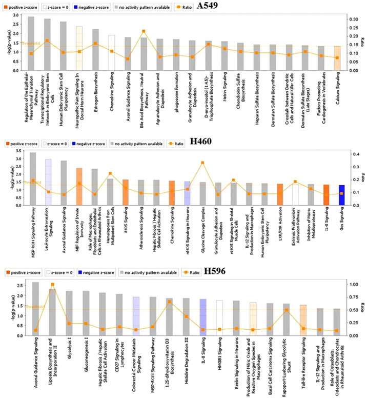
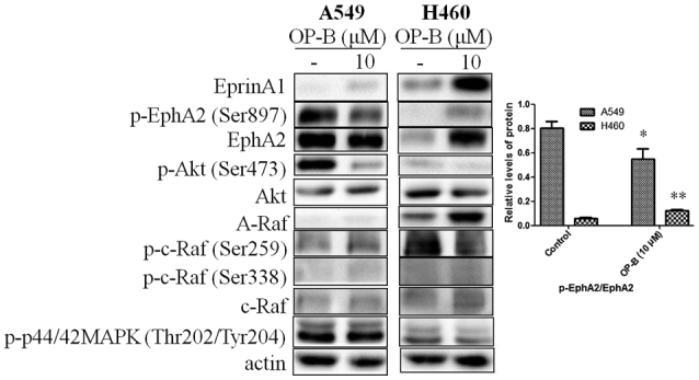
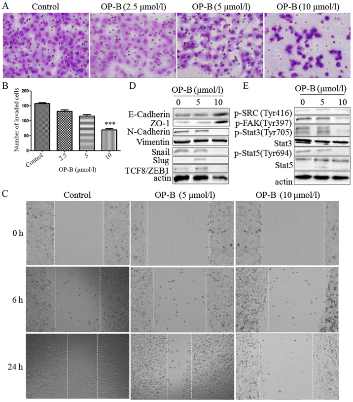
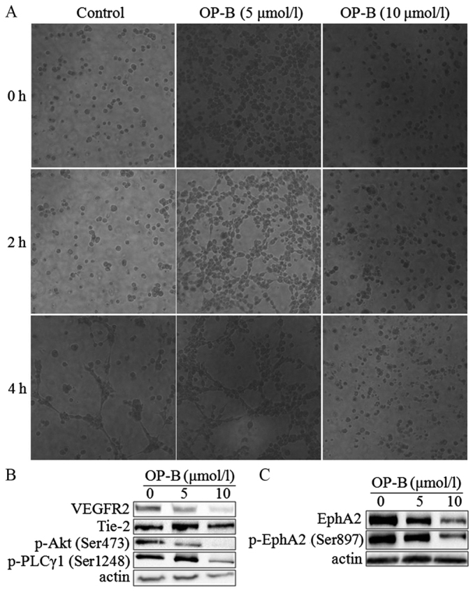
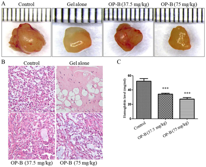
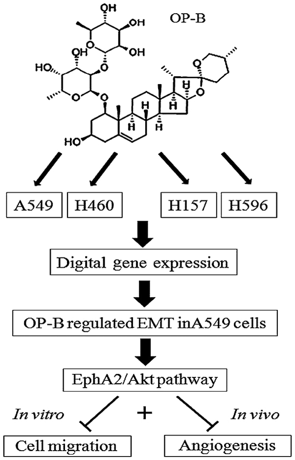
Lung adenocarcinoma is the most common metastatic cancer, and is associated with high patient mortality. Therefore, investigation of anti‑metastatic treatments for lung adenocarcinoma is crucial. Ophiopogonin B (OP‑B) is a bioactive component of Radix Ophiopogon Japonicus, which is often used in Chinese traditional medicine to treat pulmonary disease. Screening of transcriptome and digital gene expression (DGE) profiling data in NSCLC cell lines showed that OP‑B regulated the epithelial‑mesenchymal transition (EMT) pathway in A549 cells. Further results showed that 10 µmol/l OP‑B downregulated EphA2 expression and phosphorylation (Ser897) in A549 cells but upregulated them in NCI‑H460 cells. Meanwhile, the Ras/ERK pathway was unaffected in A549 cells and stimulated in NCI‑H460 cells. More importantly, detection of the EMT pathway showed that OP‑B treatment increased the epithelial markers ZO‑1 and E‑cadherin and decreased the expression of the mesenchymal marker N‑cadherin and the transcriptional repressors Snail, Slug and ZEB1. Furthermore, through Transwell migration and scratch wound healing assays, we found that 10 µmol/l OP‑B significantly reduced the invasion and migration of A549 cells. In vivo, we found that 75 mg/kg OP‑B inhibited A549 cell metastasis in a pulmonary metastasis nude mouse model. In addition, we also found that 10 µmol/l OP‑B significantly inhibited tube formation in EA.hy926 cells. The expression of VEGFR2 and Tie‑2, the phosphorylation of Akt (S473) and PLC (S1248), and the levels of EphA2 and phosphorylated EphA2 (S897) were all inhibited by OP‑B in this cell line. In vivo, using a Matrigel plug assay, we found that OP‑B inhibited angiogenesis and the hemoglobin content of A549 transplanted tumors. Taken together, OP‑B inhibited the metastasis and angiogenesis of A549 cells by inhibiting EphA2/Akt and the corresponding pathway. The investigation gives new recognition to the anticancer mechanism of OP‑B in NSCLC and this compound is a promising inhibitor of metastasis and angiogenesis of lung adenocarcinoma cells.


Figures

Figure 1.
Figure 1.
The canonical pathways in the 3 cell lines regulated by OP-B. OP-B, Ophiopogonin B.
Figure 2.
Figure 2.
Effects of OP-B on EphA2/Akt signaling pathway in A549 and NCI-H460 cells. A549 or NCI-H460 cells were treated with or without 10 µmol/l OP-B for 24 h, and then, the expression levels of EphrinA1, EphA2, A-Raf, and c-Raf and the phosphorylation levels of Akt (Ser473), EphA2 (Ser897), c-Raf (Ser259), c-Raf (Ser338), and ERK1/2 were detected by western blotting. β-actin was used as a loading control. The experiment was repeated three times and yielded similar results. Densitometric analysis of the western blots (right); n=3. *P<0.05 and **P<0.01 represent significant differences compared with control cells. OP-B, Ophiopogonin B.
Figure 3.
Figure 3.
OP-B inhibits the invasion and migration of A549 cells in vitro and in vivo. (A) Transwell migration and invasion assays were performed to examine cell migration and invasion in A549 cells. Representative images of migrated or invaded cells are displayed (magnification, ×200). (B) Columns indicate the mean ± SD of triplicate experiments (***P<0.001, independent Student's t-test). (C) Wound healing assays were used to investigate the motility of A549 cells treated with OP-B, and representative images are shown (magnification, ×40). (D and E) A549 cells were treated with or without 10 µmol/l OP-B for 24 h, and then, the expression levels of vimentin, N-cadherin, E-cadherin, ZO-1, Snail, Slug, TCF8/ZEB1, p-SRC (Tyr416), p-FAK (Tyr397), p-Stat3 (Tyr705), Stat3, p-Stat5 (Tyr694) and Stat5 were detected by western blotting. β-actin was used as a loading control. The experiment was repeated three times and yielded similar results. OP-B, Ophiopogonin B. OP-B inhibits the invasion and migration of A549 cells in vitro and in vivo. (F) OP-B inhibits the lung metastasis of A549 cells in vivo. Representative images of H&E-stained metastatic lung nodules. OP-B, Ophiopogonin B.
Figure 3.
Figure 3.
OP-B inhibits the invasion and migration of A549 cells in vitro and in vivo. (A) Transwell migration and invasion assays were performed to examine cell migration and invasion in A549 cells. Representative images of migrated or invaded cells are displayed (magnification, ×200). (B) Columns indicate the mean ± SD of triplicate experiments (***P<0.001, independent Student's t-test). (C) Wound healing assays were used to investigate the motility of A549 cells treated with OP-B, and representative images are shown (magnification, ×40). (D and E) A549 cells were treated with or without 10 µmol/l OP-B for 24 h, and then, the expression levels of vimentin, N-cadherin, E-cadherin, ZO-1, Snail, Slug, TCF8/ZEB1, p-SRC (Tyr416), p-FAK (Tyr397), p-Stat3 (Tyr705), Stat3, p-Stat5 (Tyr694) and Stat5 were detected by western blotting. β-actin was used as a loading control. The experiment was repeated three times and yielded similar results. OP-B, Ophiopogonin B. OP-B inhibits the invasion and migration of A549 cells in vitro and in vivo. (F) OP-B inhibits the lung metastasis of A549 cells in vivo. Representative images of H&E-stained metastatic lung nodules. OP-B, Ophiopogonin B.
Figure 4.
Figure 4.
OP-B inhibits tubule formation by endothelial cells. (A) Matrigel assay analysis was used to detect microtubule formation by EA.hy926 cells after treatment with 0, 5, or 10 µmol/l OP-B for 0, 2, or 4 h. Representative images are displayed (magnification, ×200). (B and C) A549 cells were treated with 0, 5 or 10 µmol/l OP-B for 24 h, and the phosphorylation levels of VEGFR2, Tie-2, p-Akt (Ser473), p-PLCγ1 (Ser1248), EphA2, and p-EphA2 (Ser897) were detected by western blotting. β-actin was used as a loading control. The experiment was repeated three times and yielded similar results. OP-B, Ophiopogonin B.
Figure 5.
Figure 5.
OP-B inhibits angiogenesis in vivo. (A and B) Tumor angiogenesis was significantly inhibited by 37.5 or 75 mg/kg OP-B. (C) The hemoglobin content of the Matrigel plugs was determined using Drabkin's reagent kit. The results are expressed as the mean ± SD (n=5) (Error bars, SD; ***P<0.001). OP-B, Ophiopogonin B.
Figure 6.
Figure 6.
The proposed mechanism of OP-B in A549 cells. Ingenuity pathway analysis of the digital gene expression indicates that OP-B specifically regulates the epithelial-mesenchymal transition pathway in A549 adenocarcinoma cells. Through inhibiting the EphA2/Akt/Raf/ERK and Src/FAK/Stat pathways in the cells, OP-B inhibits the metastasis and angiogenesis of A549 cells in vitro and in vivo. OP-B, Ophiopogonin B.

References

    1. Collins LG, Haines C, Perkel R, Enck RE. Lung cancer: Diagnosis and management. Am Fam Physician. 2007;75:56–63. - PubMed
    1. Riihimaki M, Hemminki A, Fallah M, Thomsen H, Sundquist K, Sundquist J, Hemminki K. Metastatic sites and survival in lung cancer. Lung Cancer. 2014;86:78–84. doi: 10.1016/j.lungcan.2014.07.020. - DOI - PubMed
    1. De Craene B, Berx G. Regulatory networks defining EMT during cancer initiation and progression. Nat Rev Cancer. 2013;13:97–110. doi: 10.1038/nrc3447. - DOI - PubMed
    1. Yang G, Liang Y, Zheng T, Song R, Wang J, Shi H, Sun B, Xie C, Li Y, Han J, et al. FCN2 inhibits epithelial-mesenchymal transition-induced metastasis of hepatocellular carcinoma via TGF-β/Smad signaling. Cancer Lett. 2016;378:80–86. doi: 10.1016/j.canlet.2016.05.007. - DOI - PubMed
    1. Tandon M, Vemula SV, Mittal SK. Emerging strategies for EphA2 receptor targeting for cancer therapeutics. Expert Opin Ther Targets. 2011;15:31–51. doi: 10.1517/14728222.2011.538682. - DOI - PMC - PubMed

MeSH terms