. 2018 Jun 29;14(6):e1006264.
doi: 10.1371/journal.pcbi.1006264. eCollection 2018 Jun.

Computation predicts rapidly adapting mechanotransduction currents cannot account for tactile encoding in Merkel cell-neurite complexes

Affiliations

Computation predicts rapidly adapting mechanotransduction currents cannot account for tactile encoding in Merkel cell-neurite complexes

Gregory J Gerling et al. PLoS Comput Biol. .

Abstract

Distinct firing properties among touch receptors are influenced by multiple, interworking anatomical structures. Our understanding of the functions and crosstalk of Merkel cells and their associated neurites-the end organs of slowly adapting type I (SAI) afferents-remains incomplete. Piezo2 mechanically activated channels are required both in Merkel cells and in sensory neurons for canonical SAI responses in rodents; however, a central unanswered question is how rapidly inactivating currents give rise to sustained action potential volleys in SAI afferents. The computational model herein synthesizes mechanotransduction currents originating from Merkel cells and neurites, in context of skin mechanics and neural dynamics. Its goal is to mimic distinct spike firing patterns from wildtype animals, as well as Atoh1 knockout animals that completely lack Merkel cells. The developed generator function includes a Merkel cell mechanism that represents its mechanotransduction currents and downstream voltage-activated conductances (slower decay of current) and a neurite mechanism that represents its mechanotransduction currents (faster decay of current). To mimic sustained firing in wildtype animals, a longer time constant was needed than the 200 ms observed for mechanically activated membrane depolarizations in rodent Merkel cells. One mechanism that suffices is to introduce an ultra-slowly inactivating current, with a time constant on the order of 1.7 s. This mechanism may drive the slow adaptation of the sustained response, for which the skin's viscoelastic relaxation cannot account. Positioned within the sensory neuron, this source of current reconciles the physiology and anatomical characteristics of Atoh1 knockout animals.


Conflict of interest statement

The authors have declared that no competing interests exist.

Figures
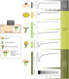
Fig 1
Fig 1. A computational model of mechanotransduction in the slowly adapting type I cutaneous afferent.
Shown is current over a ramp and hold stimulus for multiple sub-components, including a slowly inactivating (SI) current modeled as originating from the Merkel cell and rapidly inactivating (RI) and ultra-slowly inactivating (USI) currents modeled as originating from the neurite terminal. All three currents are included within the generator function, which receives input of compressive stress from a finite element model. The finite element model takes probe force, as shown in the upper panel, as its input. The generated trace of compressive stress interior to the skin’s layers, as shown in Fig 2, exhibits time-dependent viscoelastic relaxation. The currents that the generator function represents, in modeling one Merkel cell—neurite complex, are summed across a cluster of eight Merkel cell-neurite complexes feeding a heminode. It is this current, upon entering into a leaky integrate and fire model, which gives rise to predicted spike firing times. Note that for the sake of the simulation here, the irregular inter-spike intervals were unimportant so noise was removed from the model.
Fig 2
Fig 2. Conceptual view of the generator function.
The diagrams demonstrate how small time constants of about 8 and 200 ms respectively for the SI and RI components are convolved together with stress in the skin to produce a composite current from which spike firing responses are ultimately derived. In (A) stress over time, under a displacement-controlled ramp-and-hold stimulation (top bar dark grey is ramp, light grey is hold), serves as the input to the generator function. Note the viscoelastic relaxation of skin stress over the stimulus hold. Then, in three cases with inputs of step stresses, (B) a single step increase in stress σ1 evokes current output I1, which is the sum of slowly inactivating current ISI and rapidly inactivating current IRI, (C) three sequentially delivered step stresses show that current decays but builds upon the prior magnitude, and (D) a single step decrease in stress from σn to σn+1 evokes an immediate decrease in current followed by a slower decay. Note that the ultra-slowly inactivating current is omitted to simplify the explanation of the concept. Also omitted for simplicity are the finite element and leaky integrate-and-fire model contributions.
Fig 3
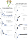
Fig 3. An ultra-slowly inactivating (USI) current is essential to drive the slow adaptation in firing in the sustained response.
In contrast, neither the modification of the SI and RI currents of generator function, nor the skin’s viscoelastic relaxation, adequately account for the slow adaptation in firing in the sustained response. Instantaneous firing frequency (IFF) plots over time are generated (panel B) similar to those observed in electrophysiological recordings (panel A). The data for wildtype animals were originally reported in Maksimovic, et. al. 2014 [3]. The two traces per plot represent two magnitudes of ramp and hold stimulation. The need for the USI component is shown in panel C. Without the USI component, the output IFF reaches a plateau and does not adapt as is typically observed for SAI afferents. Another potential way to achieve adaptation is to increase the time constant on the SI component of the model from 200 to 570 ms. However, the duration of this time constant is well outside observed bounds. For the case of the low magnitude stimulus, in panels D—F, increasing generator function parameters τRI, τSI, and KSI_Peak can increase receptor current in ramp-up, early hold, and late hold phases, respectively, as well as their corresponding IFFs (S2 Fig). They do not however impact the plateau in the sustained hold. In particular, current traces with different (D) τRI values show the impact upon the peak current produced, (E) τSI values show the impact during the early hold phase, and (F) KSI_Peak values show the impact of modulating the steady state magnitude relative to the peak. Note that in panels D—F, the USI component is included. In panel G, currents are shown that correspond to IFFs in panel C. Data similar to panel C, but for the case of high magnitude stimulation, are given in S3 Fig. The tau values are in units of ms.
Fig 4
Fig 4. Modeled response for the case of Atoh1 knockout animals.
By selectively deactivating sub-components of the generator function, IFF plots over time are generated (panel B) similar to those observed in electrophysiological recordings for Atoh1 knockout animals (panel A), in terms of attenuated temporal and spatial response compared to wildtype responses in Fig 3. Data from panel A were originally reported in Maksimovic, et. al. 2014 [3]. The two traces per plot represent two magnitudes of ramp and hold stimulation. Here, to model the Atoh1 knockout response, the SI component is removed entirely. In panel C, the current underlying the low magnitude stimulation case is shown in “Atoh1 CKO (USI and RI) Low stim.” As previously noted in panels C and G of Fig 3, an alternate approach to utilizing a USI term is to extend the duration of the SI term’s time constant. Aside from physiological feasibility, when the SI time constant is increased and USI not utilized, then the Atoh1CKO response—made up of USI and RI components—would revert to only an RI component (normalized to the magnitude of the USI and RI case) and its rate of decay does not match the Atoh1CKO current, panel C: “Atoh1 CKO (RI only, normalized).” In comparison to the electrophysiological recordings in panel A, its current decays to 0 IFF (where each line in panel C crosses the “approx. threshold for spike generation”) in 0.25 s whereas the IFF continues for 1–2 s in observed recordings. In fact, when the “Atoh1 CKO (RI only, normalized)” current is run through the full model, it produces just one spike.
Fig 5
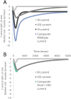
Fig 5. The respective currents underlying the composite current at one modeled Merkel cell—Neurite terminal, for wildtype (panel A) and Atoh1CKO (panel B) mouse cases.
Interestingly, across both animal types, the SI current is larger than the RI current, even in the dynamic phase of the stimulus. As well, the USI current is larger than the RI current. In the Atoh1CKO case then, it is the USI current that is nearly entirely driving the response.

Similar articles

  • Mechanotransduction in epidermal Merkel cells.
    Nakatani M, Maksimovic S, Baba Y, Lumpkin EA. Nakatani M, et al. Pflugers Arch. 2015 Jan;467(1):101-8. doi: 10.1007/s00424-014-1569-0. Epub 2014 Jul 23. Pflugers Arch. 2015. PMID: 25053537 Free PMC article. Review.
  • Piezo2 is required for Merkel-cell mechanotransduction.
    Woo SH, Ranade S, Weyer AD, Dubin AE, Baba Y, Qiu Z, Petrus M, Miyamoto T, Reddy K, Lumpkin EA, Stucky CL, Patapoutian A. Woo SH, et al. Nature. 2014 May 29;509(7502):622-6. doi: 10.1038/nature13251. Epub 2014 Apr 6. Nature. 2014. PMID: 24717433 Free PMC article.
  • Epidermal Merkel cells are mechanosensory cells that tune mammalian touch receptors.
    Maksimovic S, Nakatani M, Baba Y, Nelson AM, Marshall KL, Wellnitz SA, Firozi P, Woo SH, Ranade S, Patapoutian A, Lumpkin EA. Maksimovic S, et al. Nature. 2014 May 29;509(7502):617-21. doi: 10.1038/nature13250. Epub 2014 Apr 6. Nature. 2014. PMID: 24717432 Free PMC article.
  • Piezo2 is the major transducer of mechanical forces for touch sensation in mice.
    Ranade SS, Woo SH, Dubin AE, Moshourab RA, Wetzel C, Petrus M, Mathur J, Bégay V, Coste B, Mainquist J, Wilson AJ, Francisco AG, Reddy K, Qiu Z, Wood JN, Lewin GR, Patapoutian A. Ranade SS, et al. Nature. 2014 Dec 4;516(7529):121-5. doi: 10.1038/nature13980. Nature. 2014. PMID: 25471886 Free PMC article.
  • Merkel cells and neurons keep in touch.
    Woo SH, Lumpkin EA, Patapoutian A. Woo SH, et al. Trends Cell Biol. 2015 Feb;25(2):74-81. doi: 10.1016/j.tcb.2014.10.003. Epub 2014 Dec 2. Trends Cell Biol. 2015. PMID: 25480024 Free PMC article. Review.

Cited by

References

    1. Johnson K. The roles and functions of cutaneous mechanoreceptors. Current Opinion in Neurobiology. 2001. August 1;11(4):455–61. - PubMed
    1. Zimmerman A, Bai L, Ginty DD. The gentle touch receptors of mammalian skin. Science. 2014. November 21;346(6212):950–4. 10.1126/science.1254229 - DOI - PMC - PubMed
    1. Maksimovic S, Nakatani M, Baba Y, Nelson AM, Marshall KL, Wellnitz SA, Firozi P, Woo S-H, Ranade S, Patapoutian A, Lumpkin EA. Epidermal Merkel cells are mechanosensory cells that tune mammalian touch receptors. Nature. 2014. May 29;509(7502):617–21. 10.1038/nature13250 - DOI - PMC - PubMed
    1. Maricich SM, Wellnitz SA, Nelson AM, Lesniak DR, Gerling GJ, Lumpkin EA, Zoghbi HY. Merkel Cells Are Essential for Light-Touch Responses. Science. 2009. June 18;324(5934):1580–2. 10.1126/science.1172890 - DOI - PMC - PubMed
    1. Lesniak DR, Marshall KL, Wellnitz SA, Jenkins BA, Baba Y, Rasband MN, Gerling GJ, Lumpkin EA. Computation identifies structural features that govern neuronal firing properties in slowly adapting touch receptors. eLife. 2014. January;3:e01488 10.7554/eLife.01488 - DOI - PMC - PubMed

Publication types

LinkOut - more resources