. 2018 Jun 29;13(6):e0199312.
doi: 10.1371/journal.pone.0199312. eCollection 2018.

Enhancing radiosensitivity of melanoma cells through very high dose rate pulses released by a plasma focus device

Affiliations

Enhancing radiosensitivity of melanoma cells through very high dose rate pulses released by a plasma focus device

Francesca Buontempo et al. PLoS One. .

Erratum in

Retraction in

Abstract

Radiation therapy is a useful and standard tumor treatment strategy. Despite recent advances in delivery of ionizing radiation, survival rates for some cancer patients are still low because of recurrence and radioresistance. This is why many novel approaches have been explored to improve radiotherapy outcome. Some strategies are focused on enhancement of accuracy in ionizing radiation delivery and on the generation of greater radiation beams, for example with a higher dose rate. In the present study we proposed an in vitro research of the biological effects of very high dose rate beam on SK-Mel28 and A375, two radioresistant human melanoma cell lines. The beam was delivered by a pulsed plasma device, a "Mather type" Plasma Focus for medical applications. We hypothesized that this pulsed X-rays generator is significantly more effective to impair melanoma cells survival compared to conventional X-ray tube. Very high dose rate treatments were able to reduce clonogenic efficiency of SK-Mel28 and A375 more than the X-ray tube and to induce a greater, less easy-to-repair DNA double-strand breaks. Very little is known about biological consequences of such dose rate. Our characterization is preliminary but is the first step toward future clinical considerations.


Conflict of interest statement

The authors have declared that no competing interests exist.

Figures

Fig 1
Fig 1. PFMA-3 causes radiation-induced clonogenic cell death greater than XRT.
(a) Colony forming unit assay on SK-Mel28 and A375 cells irradiated with PFMA-3 and XRT at 2, 4 and 8 Gy. Data are representative of three independent experiments performed in triplicate and SD is not shown being less than 10%. Black (SK-Mel28) and red (A375) asterisks indicate statistically significant differences (*P<0.05, **P <0.005). (b) Radiobiological parameters of SK-Mel28 and A375 cell lines for PFMA-3 and XRT 2, 4, 8 Gy irradiation. SF2, SF4 and SF8 survival fraction at 2, 4 and 8 Gy respectively. D50, 50% survival dose. The D50 values for XRT device were used as standard reference for the RBE evaluations. The MLQ curve has been built from parameters suggested for SK-Mel28 and for low energy X-ray sources with a correction for the exposure time [37].
Fig 2
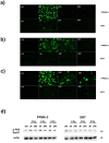
Fig 2. PFMA-3 causes a DNA-DSB more severe with respect to XRT in SK-Mel28.
Induction of DSB was assessed through detection of phosphorylation of H2A.X at Ser139 (green) by immunofluorescence and microscopy analysis. a) DSB after 2 Gy treatment. b) DSB after 4 Gy treatment. c) DSB after 8 Gy treatment. ctrl, control cells. d) Western blot analysis documenting phospho-p53 (Ser51) levels modulation in PFMA-3 and XRT irradiated samples. Antibody to b-actin served as a loading control. ctrl, control cells.
Fig 3
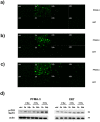
Fig 3. PFMA-3 causes a DNA-DSB more severe with respect to XRT in A375.
Induction of DSB was assessed through detection of phosphorylation of H2A.X at Ser139 (green) by immunofluorescence and microscopy analysis. a) DSB after 2 Gy treatment. b) DSB after 4 Gy treatment. c) DSB after 8 Gy treatment. ctrl, control cells. d) Western blot analysis documenting phospho-p53 (Ser51) levels modulation in PFMA-3 and XRT irradiated samples. Antibody to b-actin served as a loading control. ctrl, control cells.
Fig 4
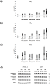
Fig 4. PFMA-3 and XRT treatments did not induce significant apoptotic events until 96 hours.
Flow cytometric analysis of Annexin V-FITC/PI-stained SK-Mel28 cells treated with a) 2 Gy b) 4 Gy c) 8 Gy PFMA-3 and XRT at different time points. The percentages of early apoptotic cells (Annexin V FITC+/PI-), late apoptotic/necrotic cells (Annexin V FITC+/PI+) and necrotic cells (Annexin V FITC-/PI+) were plotted. The histograms were representative of three separate experiments. Asterisks indicated statistically significant differences (**P <0.005). ctrl, control cells. d) Western blot analysis documenting some apoptotic marker levels modulation in PFMA-3 and XRT irradiated samples. Antibody to b-actin served as a loading control. ctrl, control cells.
Fig 5
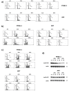
Fig 5. PFMA-3 very high dose rate altered SK-Mel28 cell cycle distribution more severely than XRT and finally induced the appearance of sub-G1 peak.
Flow cytometric analysis of cell cycle distribution in SK-Mel28 cell line treated with a) 2 Gy b) 4 Gy c) 8 Gy PFMA-3 and XRT for different times. The histograms were representative of three separate experiments. ctrl, control cells. d) Western blot analysis documenting cyclin-B1 levels modulation in PFMA-3 and XRT irradiated cells. Antibody to b-actin served as a loading control. ctrl, control cells.
Fig 6
Fig 6. PFMA-3 caused higher levels of SIPS respect to XRT.
Representative images of staining for senescence-associated β-galactosidase 96 hrs following irradiation with 2, 4 and 8 Gy PFMA-3 and XRT. Data were representative of three independent experiments. ctrl, control cells.
Fig 7
Fig 7. PFMA-3 affected SK-Mel28 migration abilities more than XRT.
a) Cell migration behaviour was evaluated during performance of a wound-healing assay after treatment with 2 Gy, 4 Gy and 8 Gy PFMA-3 and XRT. Data were representative of three independent experiments performed in quadruplicate. ctrl, control cells. Asterisks indicated statistically significant differences (*P <0.05, **P <0.005, ***P <0.0005). b) Western blot analysis documenting E-cadherin levels modulation in PFMA-3 and XRT irradiated samples. b-actin loading control was not shown. ctrl, control cells.
Fig 8
Fig 8. PFMA-3 affected A375 migration abilities more than XRT.
Cell migration behaviour was evaluated during performance of a wound-healing assay after treatment with 2 Gy, 4 Gy and 8 Gy PFMA-3 and XRT. Data were representative of three independent experiments performed in quadruplicate. ctrl, control cells. Asterisks indicated statistically significant differences (*P <0.05, **P <0.005, ***P <0.0005).
Fig 9
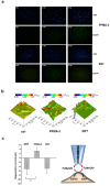
Fig 9. PFMA-3 induced glutathione and lipid peroxidation production greater than XRT.
a) Visualization of the SK-Mel28 living cells 3 hrs after irradiation with PFMA-3 or XRT (at 2 and 4 Gy) and of those non irradiated (ctrl) loaded by the fluorescent dyes monochloromobimane (mBCl) and boron-dipyrromethene (BODIPY) for a qualitative GSH and lipid peroxidation detection, respectively. Epifluorescence images at 100 × of magnification. b) Representative FcMeOH/SECM images in constant height mode of non irradiated (ctrl) and irradiated SK-Mel28 cells at 4 Gy by PFMA-3 and XRT. The regeneration currents recorded were reported as a percentage increase/decrease of the signal in respect to the one recorded at the same tip-dish distance but far away from cells (IT/ITdish). c) Histogram reported the values obtained by computing the mean (± SEM) regeneration current recorded on 4 different cells selected from the scanning electrochemical microscopy images. Asterisks indicated statistically significant differences (*P <0.05).

Similar articles

Cited by

References

    1. Delaney G, Jacob S, Featherstone C, Barton M. The role of radiotherapy in cancer treatment: estimating optimal utilization from a review of evidence-based clinical guidelines. Cancer. 2005;104(6):1129–37. doi: 10.1002/cncr.21324 . - DOI - PubMed
    1. Bhide SA, Nutting CM. Recent advances in radiotherapy. BMC Med. 2010;8:25 doi: 10.1186/1741-7015-8-25 . - DOI - PMC - PubMed
    1. Baumann M, Krause M, Overgaard J, Debus J, Bentzen SM, Daartz J, et al. Radiation oncology in the era of precision medicine. Nat Rev Cancer. 2016;16(4):234–49. doi: 10.1038/nrc.2016.18 . - DOI - PubMed
    1. Maier P, Hartmann L, Wenz F, Herskind C. Cellular Pathways in Response to Ionizing Radiation and Their Targetability for Tumor Radiosensitization. Int J Mol Sci. 2016;17(1). doi: 10.3390/ijms17010102 . - DOI - PMC - PubMed
    1. Bentzen SM, Ritter MA. The alpha/beta ratio for prostate cancer: what is it, really? Radiother Oncol. 2005;76(1):1–3. doi: 10.1016/j.radonc.2005.06.009 . - DOI - PubMed

Publication types