Comparative Study
. 2018 Sep 4;26(9):1169-1177.e3.
doi: 10.1016/j.str.2018.05.006. Epub 2018 Jun 26.

Refinement and Analysis of the Mature Zika Virus Cryo-EM Structure at 3.1 Å Resolution

Affiliations
Comparative Study

Refinement and Analysis of the Mature Zika Virus Cryo-EM Structure at 3.1 Å Resolution

Madhumati Sevvana et al. Structure. .

Abstract

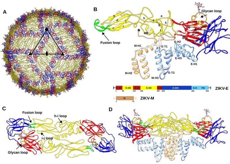
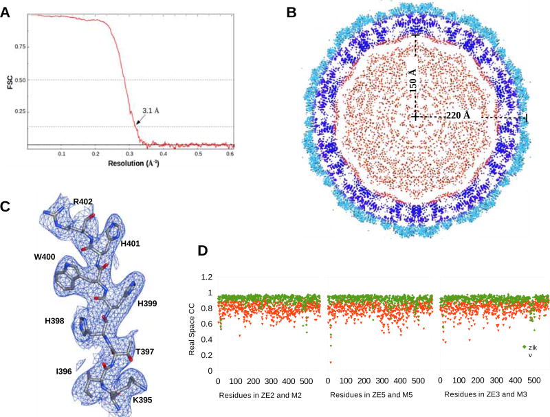
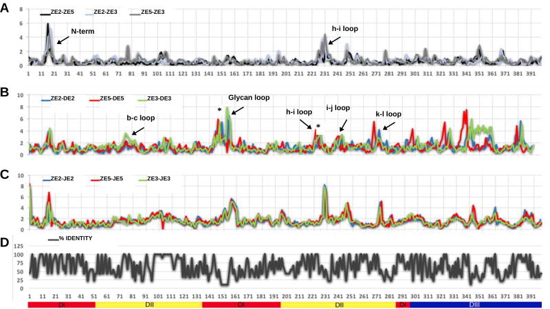
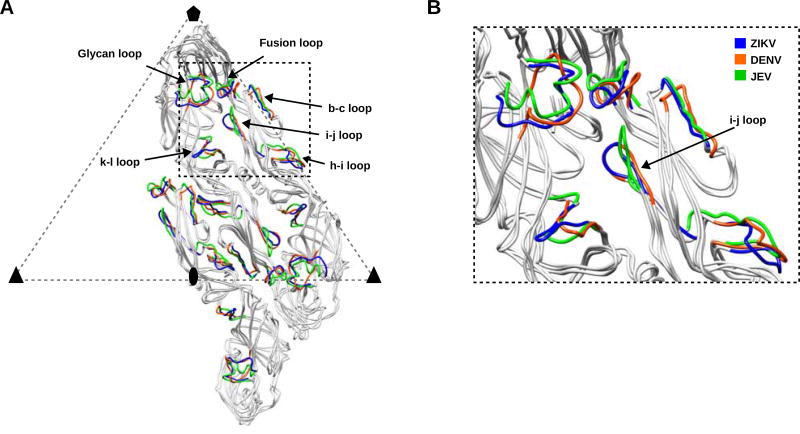
Among the several arthropod-borne human flaviviral diseases, the recent outbreak of Zika virus (ZIKV) has caused devastating birth defects and neurological disorders, challenging the world with another major public health concern. We report here the refined structure of the mature ZIKV at a resolution of 3.1 Å as determined by cryo-electron microscopic single-particle reconstruction. The improvement in the resolution, compared with previous enveloped virus structures, was the result of optimized virus preparation methods and data processing techniques. The glycoprotein interactions and surface properties of ZIKV were compared with other mosquito-borne flavivirus structures. The largest structural differences and sequence variations occur at the glycosylation loop associated with receptor binding. Probable drug binding pockets were identified on the viral surface. These results also provide a structural basis for the design of vaccines against ZIKV.

Keywords: Zika virus; antiviral binding sites; flavivirus comparisons; receptor binding sites; structure refinement.


Conflict of interest statement

DECLARATION OF INTERESTS

The authors declare no competing financial interests.

Figures

Figure 1
Figure 1. Overall Structure of ZIKV
(A) ZIKV structure highlighting the herringbone pattern formed by 6 E–M heterodimers. The icosahedral asymmetric unit, is outlined by a black triangle. The structures of the E proteins are shown as Cα backbone traces. Domains E-DI, E-DII and E-DIII of each E protein are shown in red, yellow and blue, respectively. The fusion loop of each E protein molecule is shown in green. (B) Secondary structural elements of one E–M heterodimer are labelled. Secondary structure elements on E-DI, E-DII and E-DIII are labelled A0–I0, a–l and A–G, respectively. The stem and transmembrane helices (E-H1, E-H2, E-H3, E-T1, E-T2) of the E protein and the M protein (M-H1, M-H2, M-H3) are colored in light blue and light brown, respectively. Residue numbering and domain definitions are shown as a linear peptide. Domains E-DI, E-DII and E-DIII are colored as in Figure 1A. Panels (C) and (D) show the top and side views of the dimer, respectively. The glycan loop, fusion loop, h–I loop and i–j loop are labelled. (See also Table S1.)
Figure 2
Figure 2. The Cryo-EM Reconstruction of ZIKV
(A) Fourier Shell Coefficient plot versus resolution. (B) A central section looking down a 5-fold axis of the cryo-EM electron potential map. The E-ectodomain (1–400), the E and M transmembrane regions and the core are colored light blue, dark blue and red, respectively. (C) The cryo-EM map contoured at 1.5 σ around residues Lys395-Arg402. (D) Plot showing real space correlation coefficient for the residues in the current structure (green) and the earlier structure (PDB ID:5IRE) (red).
Figure 3
Figure 3. Comparison of ZIKV, DENV and JEV Structures
(A) R.m.s.d between equivalent Cα atoms of the three independent monomers in ZIKV (ZE2, ZE3, ZE5). (B) R.m.s.d between equivalent Cα atoms of DENV monomers (DE2, DE3, DE5) and ZIKV E monomers. (C) R.m.s.d between equivalent Cα atoms of JEV (JE2, JE3, JE5) monomers and ZIKV monomers. (D) Plot showing percent sequence identity from multiple sequence alignment as in Figure S1. The secondary structure elements with larger r.m.s.d’s are labeled in panels (A) and (B). Sequence insertions on ZIKV are marked with an asterisk on panel (B). In (A), (B) and (C) the x-axis shows amino acid residue numbering and the y-axis shows the r.m.s.d between Cα atoms. The secondary structure elements of the E-ectodomain are labelled below panel (D). (See also Figures S1 and S2.)
Figure 4
Figure 4. Superposition of Cα Atoms of the E Monomers in One Asymmetric Unit of DENV and JEV on ZIKV
(A) The E2, E3 and E5 monomers of ZIKV, DENV and JEV are colored in grey ribbons. Structural elements with maximum r.m.s.d’s in ZIKV, DENV and JEV are colored in blue, orange and green, respectively. (B) The inset shows a region around the glycan loop and the differences in closed and open conformations of i–j loop in DENV and JEV respectively.
Figure 5
Figure 5. Surface-Exposed Residues in ZIKV, DENV and JEV
Roadmaps of surface-exposed residues in ZIKV (panels A and B), DENV (panels C and D) and JEV (panels E and F). Panels B, D, and F depict regions near the glycan loop as outlined by the white rectangles in panels A, C, and E, respectively. In (B) ZIKV and (F) JEV, the region formed by the E-DIII surface residues and the adjacent glycan loop (shown in red) is rich in serine and threonine residues, whereas (D) DENV has a different residue distribution in the same region. The shape of the motif formed by the surface exposed residues is similar in (B) ZIKV and (F) JEV. (See also Figures S3, S4, S5 and Tables S2, S3 and S4.)

Similar articles

Cited by

References

    1. Afonine PV, Grosse-Kunstleve RW, Echols N, Headd JJ, Moriarty NW, Mustyakimov M, Terwilliger TC, Urzhumtsev A, Zwart PH, Adams PD. Towards automated crystallographic structure refinement with phenix.refine. Acta Crystallogr. D Biol. Crystallogr. 2012;68:352–367. - PMC - PubMed
    1. Alhoot MA, Rathinam AK, Wang SM, Manikam R, Sekaran SD. Inhibition of dengue virus entry into target cells using synthetic antiviral peptides. Int. J. Med. Sci. 2013;10:719–729. - PMC - PubMed
    1. Annamalai AS, Pattnaik A, Sahoo BR, Muthukrishnan E, Natarajan SK, Steffen D, Vu HLX, Delhon G, Osorio FA, Petro TM, et al. Zika virus encoding non-glycosylated envelope protein is attenuated and defective in neuroinvasion. J. Virol. 2017;91:e01348–01317. - PMC - PubMed
    1. Barba-Spaeth G, Dejnirattisai W, Rouvinski A, Vaney MC, Medits I, Sharma A, Simon-Loriere E, Sakuntabhai A, Cao-Lormeau VM, Haouz A, et al. Structural basis of potent Zika-dengue virus antibody cross-neutralization. Nature. 2016;536:48–53. - PubMed
    1. Brasil P, Pereira JP, Moreira ME, Nogueira RMR, Damasceno L, Wakimoto M, Rabello RS, Valderramos SG, Halai UA, Salles TS, et al. Zika virus infection in pregnant women in Rio de Janeiro. N. Engl. J. Med. 2016;375:2321–2334. - PMC - PubMed

Publication types