. 2018 Jul 17;49(1):56-65.e4.
doi: 10.1016/j.immuni.2018.04.032. Epub 2018 Jun 26.

The TWIK2 Potassium Efflux Channel in Macrophages Mediates NLRP3 Inflammasome-Induced Inflammation

Affiliations

The TWIK2 Potassium Efflux Channel in Macrophages Mediates NLRP3 Inflammasome-Induced Inflammation

Anke Di et al. Immunity. .

Abstract

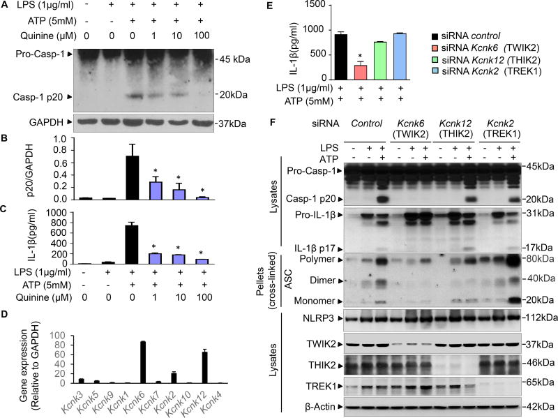
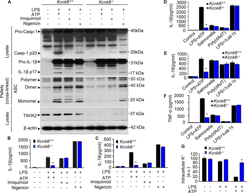
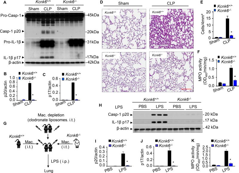
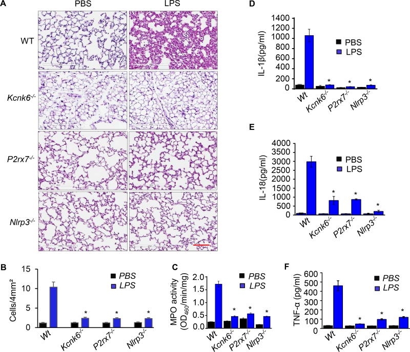
Potassium (K+) efflux across the plasma membrane is thought to be an essential mechanism for ATP-induced NLRP3 inflammasome activation, yet the identity of the efflux channel has remained elusive. Here we identified the two-pore domain K+ channel (K2P) TWIK2 as the K+ efflux channel triggering NLRP3 inflammasome activation. Deletion of Kcnk6 (encoding TWIK2) prevented NLRP3 activation in macrophages and suppressed sepsis-induced lung inflammation. Adoptive transfer of Kcnk6-/- macrophages into mouse airways after macrophage depletion also prevented inflammatory lung injury. The K+ efflux channel TWIK2 in macrophages has a fundamental role in activating the NLRP3 inflammasome and consequently mediates inflammation, pointing to TWIK2 as a potential target for anti-inflammatory therapies.

Keywords: KCNK6; NLRP3 inflammasome; P2X7 receptor; TWIK2; inflammation; potassium channel.


Conflict of interest statement

DECLARATION OF INTERESTS

The authors declare no competing interests.

Figures

Figure 1
Figure 1. TWIK2 mediated quinine sensitive NLRP3 inflammasome activation in macrophages
AC: Quinine inhibits ATP-induced NLRP3 inflammasome activation. Mouse bone marrow derived macrophages (BMDMs) were primed with LPS for 3h and subsequently challenged with ATP for 30min. Cell lysates were immunoblotted with caspase-1 p20 antibody (A) and results are summarized in B (* p < 0.05 compared with group treated with LPS and ATP but without quinine, n = 3). IL-1β release in supernatants was measured using ELISA method (C, * p < 0.05 compared with group treated with LPS and ATP but without quinine, n = 3). D: mRNA of various two-pore domain K channel (K2P) in BMDMs were assessed by qRT-PCR for the following: Kcnk1 (encoding TWIK1), Kcnk6 (encoding TWIK2), Kcnk7 (encoding TWIK3); TWIK1, TWIK2, TWIK3: 2 pore domain weak inwardly rectifying K+ channel 1, 2, and 3; Kcnk3 (encoding TASK1), Kcnk5 (encoding TASK2), Kcnk9 (encoding TASK3); TASK1, TASK2, TASK3: TWIK related acid-sensitive K+ channel 1, 2, and 3; Kcnk2 (encodingTREK1), Kcnk10 (encodingTREK2); TREK1, TREK2: TWIK related K+ channel 1 and 2; Kcnk12 (encodingTHIK2: 2 pore domain halothane-inhibited K+ channel 2); Kcnk4 (encoding TRAAK: TWIK related arachidonic acid-stimulated K+ channel). Kcnk6, Kcnk2 and Kcnk12 exhibited the highest expression (n = 3). EF: TWIK2 silenced by siRNA inhibited ATP-induced NLRP3 inflammasome activation. Mouse J774A.1 (cell line) MΦs were treated with control siRNA or siRNA targeting Kcnk6, Kcnk2 and Kcnk12, respectively. IL-1β release in supernatants was monitored with ELISA method (E, * p < 0.05 compared with siRNA control group, n = 3) and cell lysates or pellets were immunoblotted with indicated antibodies (F, representative of 3 independent experiments). NLRP3: nucleotide-binding oligomerization domain-like receptor (NLR) containing pyrin domain 3; ASC: apoptosis-associated speck-like protein containing a carboxy-terminal caspase recruitment domain (CARD). Please also see Figure S1, S2, S3 and S4.
Figure 2
Figure 2. TWIK2 mediated and P2X7 independent K+ efflux in macrophages
AF: Representative current traces at ramp voltages of whole cell patch clamp recordings with time in BMDMs from wild type (WT, A and B, 7 independent experiments), P2rx7−/− (C and D, 5 independent experiments) and Kcnk6−/− (E and F, 6 independent experiments) mice. Cells were bathed in solutions with K+ as the major outward current and Na+ and Ca2+ as the major inward current. Whole cell current was elicited with ramp voltages running from −110 mV to +50 mV within 200 ms were applied to cells with an interval of 1s. The direction of currents was indicated by straight black arrows. The “increased phase” means the currents increase with time from basal level to the peak of positive (outward) current or to the peak of negative (inward) current (indicated by color arrows). The “decreased phase” means the currents decrease with time from the peak of the outward currents or from the peak of inward) current to the basal level (indicated by color arrows); GH: Comparison of basal and ATP-induced peak current in the absence of quinine (G as described in A) and of ATP-induced peak current in the presence of quinine (H, representative of 3 independent experiments) in the different cells as indicated. The amplitudes of the outward peak currents (under 50 mV) and inward peak current (under −100 mV) from experiments described in AF and H are summarized in I (* p < 0.05 compared with WT group in each condition). Please also see Figure S2, S3, S4 and Figure S5.
Figure 3
Figure 3. TWIK2 mediated K+ efflux is required for NLRP3 inflammasome activation
AC. TWIK2 deletion (Kcnk6−/−) prevented ATP induced NLRP3 inflammasome activation in BMDMs but had no effect on neither imiquimod nor nigericin activated responses. Cells were primed with LPS (3h) and subsequently challenged with ATP (5 mM) or Imiquimod (10 µg/ml) or Nigericin (10 µg/ml) for 30 min. Cell lysates or pellets were immunoblotted with indicated antibodies (A, representative results of 3 independent experiments). IL-1β and IL-18 release in supernatants was measured using ELISA method (B and C, * p < 0.05 compared with Kcnk6+/+ plus LPS and ATP group, n = 3). DF. Kcnk6 deletion prevented NLLRP3 (not NLRC4, AIM2, or pyrin) inflammasome activation in BMDMs. BMDMs from Kcnk6+/+ and Kcnk6−/− mice were either used in an unprimed state or primed with LPS and then challenged with ATP (5mM) for 30 min or Salmonella (5 moi, to activate NLRC4) for 2h or poly (dAdT), to activate AIM2) (2 µg/ml) for 2h or TcdB toxin (0.5 µg/ml, to activate pyrin) for 3h. IL-1β (D), IL-18 (E) and TNF-α (F) concentrations in cell-free supernatants were quantified by ELISA. NLRC4: NLR family, CARD domain containing 4; AIM2: absent in melanoma 2. * p < 0.05 compared with Kcnk6+/+ plus LPS and ATP group, n = 3. G. TWIK2 deletion prevented ATP-induced decrease in intracellular potassium (K+) concentration. BMDMs were primed with LPS (3h) and then treated with ATP (30min). Intracellular K+ was examined by FluxOR™ II green potassium ion channel assay. * p < 0.05 compared with Kcnk6+/+ plus LPS and ATP group, n = 6. Please also see Figure S5 D – E and Figure S6 A – B.
Figure 4
Figure 4. TWIK2 deficiency prevents sepsis-induced NLRP3 inflammasome activation and inflammatory lung injury in mice
AF: Deletion of Kcnk6 (encoding TWIK2) prevents sepsis-induced NLRP3 inflammasome activation and inflammatory lung injury in mice. Murine lung inflammatory injury resulting from polymicrobial sepsis was induced through cecal ligation puncture (CLP) in both Kcnk6+/+ and Kcnk6−/− mice. NLRP3 inflammasome activation (indicated by caspase 1 activation and IL-1β maturation) in the murine lung was assessed by Immunoblotting (A) and quantified in B and C (* p < 0.05 compared with Kcnk6+/+ CLP group, n = 3). Representative H&E images of lung sections from 3 independent experiments are shown in D. Scale bars: 200 µm. Lung injury shown in D was evaluated by quantification of inflammatory cells in alveoli (per 4mm2 using the Fiji image analysis software) in E, and MPO measurements of lung tissue are shown in F (* p < 0.05 compared with Kcnk6+/+ CLP group, n = 3). GK: Kcnk6 deletion in macrophages prevents sepsis-induced NLRP3 inflammasome activation and inflammatory lung injury in mice. Lung macrophages (Mac) were depleted with clodronate liposomes and then reconstituted intratracheally with BMDMs either from Kcnk6+/+ or Kcnk6−/− mice as illustrated in G. The mice were injected with LPS (intra-peritoneal injection, i.p.) after 24 h of macrophage reconstitution. Lungs were harvested for evaluation of NLRP3 inflammasome activation (HJ) and neutrophil infiltration by checking MPO concentration (K), * p < 0.05 compared with Kcnk6+/+ LPS group, n = 3. Please also see Figure S6C.
Figure 5
Figure 5. Prevention of LPS-induced inflammation by TWIK2 deletion is mimicked by deleting P2X7 or NLRP3 in mice
AC: Prevention of LPS-induced lung inflammation by TWIK2 deficiency renders similar responses to LPS in P2X7 or NLRP3 deletion mice. Mouse lung inflammation was induced by intra-peritoneal injection (i.p.) of LPS and histological sections of lungs from wild type (WT), TWIK2 deficiency (Kcnk6−/−), P2X7 deficiency (P2rx7−/−) and NLRP3 deficiency (Nlrp3−/−) were stained with hematoxylin and eosin (HE) 24h of LPS injection to assess immune cell infiltration (A, representative results of 3 independent experiments; scale bars = 100 µm). Lung injury was evaluated by inflammatory cell quantification in alveoli per 4mm2 using the Fiji image analysis software (B) and lung myeloperoxidase (MPO) activity (C). * p < 0.05 compared with WT LPS group, n = 3. DF: TWIK2 deficiency prevented LPS-induced cytokine increases in serum which was phenocopied in P2rx7−/− or Nlrp3−/− mice. Concentrations of IL-1β, IL −18, and TNF-α in the mice mentioned in AC were evaluated with ELISA methods. * p < 0.05 compared with WT LPS group, n = 3.
Figure 6
Figure 6. P2X7 mediated influx of cations functions in cooperation with K+ efflux through TWIK2 to induce NLRP3 inflammasome activation in macrophages
A: TWIK2 deficiency prevents LPS-induced NLRP3 inflammasome activation similar to what is observed with P2X7 or NLRP3 deficiency in mice. Mouse lung inflammasome activation was induced by intra-peritoneal injection (i.p.) of LPS for 1 day. NLRP3 inflammasome activation (as indicated by caspase-1 activation and IL-1β maturation) was assessed in murine lungs using immunoblotting. Representative results from 3 independent experiments are shown. B: TWIK2 deficiency (Kcnk6−/−) prevented LPS-primed and ATP-induced NLRP3 inflammasome activation, and this response was similar what was seen in P2X7 deficient (P2rx7−/−) and caspase 1 deficient (Casp1−/−) cells (BMDMs). * p < 0.05 compared with WT plus LPS and ATP group, n = 3. C: Depletion of extracellular calcium (Ca2+) alone or sodium (Na+) alone only partly inhibited ATP-induced NLRP3 inflammasome activation. BMDMs were primed with LPS and then stimulated with ATP at different extracellular concentration of K+, Ca2+ and Na+ as indicated. * p < 0.05 compared with LPS plus ATP but without extracellular K+ ([K+]O = 0), n = 3 – 12. IL-1β release in BMDMs was evaluated by ELISA method.

References

    1. Achan J, Talisuna AO, Erhart A, Yeka A, Tibenderana JK, Baliraine FN, Rosenthal PJ, D'Alessandro U. Quinine, an old anti-malarial drug in a modern world: role in the treatment of malaria. Malaria journal. 2011;10:144. - PMC - PubMed
    1. Bachmaier K, Toya S, Gao X, Triantafillou T, Garrean S, Park GY, Frey RS, Vogel S, Minshall R, Christman JW, et al. E3 ubiquitin ligase Cblb regulates the acute inflammatory response underlying lung injury. Nat Med. 2007;13:920–926. - PubMed
    1. Bobak N, Feliciangeli S, Chen CC, Ben Soussia I, Bittner S, Pagnotta S, Ruck T, Biel M, Wahl-Schott C, Grimm C, et al. Recombinant tandem of pore-domains in a Weakly Inward rectifying K+ channel 2 (TWIK2) forms active lysosomal channels. Scientific reports. 2017;7:649. - PMC - PubMed
    1. Broz P, Dixit VM. Inflammasomes: mechanism of assembly, regulation and signalling. Nat Rev Immunol. 2016;16:407–420. - PubMed
    1. Buell G, Lewis C, Collo G, North RA, Surprenant A. An antagonist-insensitive P2x receptor expressed in epithelia and brain. EMBO J. 1996;15:55–62. - PMC - PubMed

Publication types

MeSH terms