Review
. 2018 Aug;29(8):2039-2052.
doi: 10.1681/ASN.2018050488. Epub 2018 Jun 29.

Nephrotoxicity of Cancer Immunotherapies: Past, Present and Future

Affiliations
Review

Nephrotoxicity of Cancer Immunotherapies: Past, Present and Future

Mark A Perazella et al. J Am Soc Nephrol. 2018 Aug.

Abstract

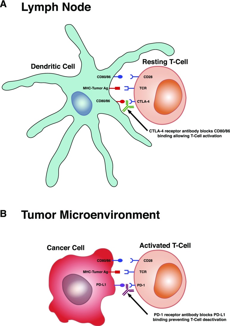
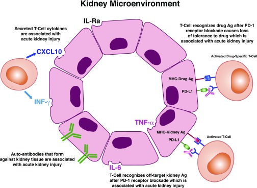
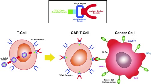
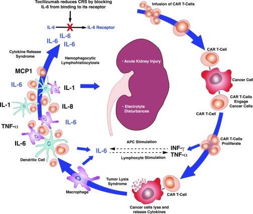
Nephrotoxicity from cancer therapies is common and increasingly encountered in clinical practice, such that the subfield of "onco-nephrology" has emerged. Conventional chemotherapeutic drugs and novel agents targeting specific genes/proteins are effective cancer therapies but suffer from a number of adverse kidney effects. An effective avenue of cancer treatment is immunotherapy, which uses drugs that augment immune system-mediated recognition and targeting of tumor cells. As such, leveraging the immune system to target malignant cells represents an important modality in eradicating cancer. IFN and high-dose IL-2 are older immunotherapies used in clinical practice to treat various malignancies, whereas new cancer immunotherapies have emerged over the past decade that offer even more effective treatment options. The immune checkpoint inhibitors are an exciting addition to the cancer immunotherapy armamentarium. Chimeric antigen receptor T cells are also a new immunotherapy used to treat various hematologic malignancies. However, as with the conventional and targeted cancer agents, the immunotherapies are also associated with immune-related adverse effects, which includes nephrotoxicity.

Keywords: acute kidney injury; chimeric antigen receptor T-cells; immune checkpoint inhibitors; immunotherapies; interferon; interleukin-2.


Figures

Figure 1.
Figure 1.
Immune checkpoint inhibitor target sites. (A) At the level of the lymph node, T cells express cytotoxic lymphocyte–associated antigen-4 (CTLA-4) receptor, which competes with CD28 receptor binding to CD80/86 on dendritic cells. This competitive binding reduces the magnitude of the CD28 costimulatory response, thus inhibiting T cell activation. This maintains balance to prevent T cell activation against self-antigen, thereby avoiding autoimmunity. Blocking CD80/86 binding to the CTLA-4 receptor with the mAb ipilimumab allows CD28 receptor binding to fully activate the T cells. This is appropriate for activation against presented tumor antigen (Ag). (B) In the tumor microenvironment, programmed death-1 (PD-1) receptor is a protein expressed on activated T cells. Certain tumors express ligands (programmed death ligand-1 [PD-L1] and programmed death ligand-2 [PD-L2]) that bind T cell PD-1 receptors and blunt T cell function. Blocking tumor cell PD-L1 or PD-L2 binding to T cell PD-1 receptor with the mAbs nivolumab and pembrolizumab leads to further T cell activation (or blocks deactivation of T cells) and promotes tumor cell killing. TCR, T cell receptor.
Figure 2.
Figure 2.
Potential mechanisms underlying immune checkpoint inhibitor–induced kidney injury. Formation of new or reactivated T cells against tumor antigens (Ags) that crossreact with off-target kidney tissues, loss of tolerance with reactivation of drug-specific T cells induced by the immune checkpoint inhibitors, increase in proinflammatory cytokines/chemokines in kidney tissue, and generation of autoantibodies (antikidney tissue antibodies) are potential mechanisms of immune checkpoint–induced kidney injury. CXCL10, C-X-C motif chemokine ligand 10; PD-1, programmed death-1; PD-L1, programmed death ligand-1.
Figure 3.
Figure 3.
Chimeric antigen receptor (CAR) T cells. T cells harvested from patients are genetically modified using lentiviral vector to place an antigen binding domain (recognizes tumor antigen), which is linked to an intracellular costimulatory domain (CD28 or 4–1BB) and CD3-ζ signaling domain to amplify the immune response against tumor cells. CAR T cells engage tumor antigen by using extracellular antigen receptors, which are linked to intracellular costimulatory and signaling domains to amplify the immune response against tumor cells. Proinflammatory cytokines and chemokines are produced, which participate in eradication of cancer cells. CXCL10, C-X-C motif chemokine ligand 10.
Figure 4.
Figure 4.
Chimeric antigen receptor (CAR) T cell infusion–mediated toxicity. CAR T cells are infused and then interact with cancer cells, where they expand further and release IFN-γ and TNF-α. These stimulate macrophage activation. CAR T cells also cause lysis of cancer cells, which leads to release of cytokines and activation of macrophages and dendritic cells. IL-1, IL-6, and IL-8 are released as well as TNF-α and monocyte chemo-attractant 1 (MCP1). IL-6 is the most significant cytokine in the cytokine release syndrome. AKI and electrolyte abnormalities may occur in this setting. Tocilizumab blocks IL-6 from binding to its receptor, reducing the effects of cytokine release syndrome (CRS). APC, antigen-presenting cell.

Similar articles

Cited by

References

    1. Rosner MH, Perazella MA: Acute kidney injury in patients with cancer. N Engl J Med 376: 1770–1781, 2017 - PubMed
    1. Porta C, Cosmai L, Gallieni M, Pedrazzoli P, Malberti F: Renal effects of targeted anticancer therapies. Nat Rev Nephrol 11: 354–370, 2015 - PubMed
    1. Schreiber RD, Old LJ, Smyth MJ: Cancer immunoediting: Integrating immunity’s roles in cancer suppression and promotion. Science 331: 1565–1570, 2011 - PubMed
    1. Khalil DN, Smith EL, Brentjens RJ, Wolchok JD: The future of cancer treatment: Immunomodulation, CARs and combination immunotherapy. Nat Rev Clin Oncol 13: 273–290, 2016 - PMC - PubMed
    1. Hanahan D, Weinberg RA: Hallmarks of cancer: The next generation. Cell 144: 646–674, 2011 - PubMed