. 2018 Aug;29(8):2123-2138.
doi: 10.1681/ASN.2017121312. Epub 2018 Jun 29.

GAPVD1 and ANKFY1 Mutations Implicate RAB5 Regulation in Nephrotic Syndrome

Affiliations

GAPVD1 and ANKFY1 Mutations Implicate RAB5 Regulation in Nephrotic Syndrome

Tobias Hermle et al. J Am Soc Nephrol. 2018 Aug.

Abstract

Background: Steroid-resistant nephrotic syndrome (SRNS) is a frequent cause of CKD. The discovery of monogenic causes of SRNS has revealed specific pathogenetic pathways, but these monogenic causes do not explain all cases of SRNS.

Methods: To identify novel monogenic causes of SRNS, we screened 665 patients by whole-exome sequencing. We then evaluated the in vitro functional significance of two genes and the mutations therein that we discovered through this sequencing and conducted complementary studies in podocyte-like Drosophila nephrocytes.

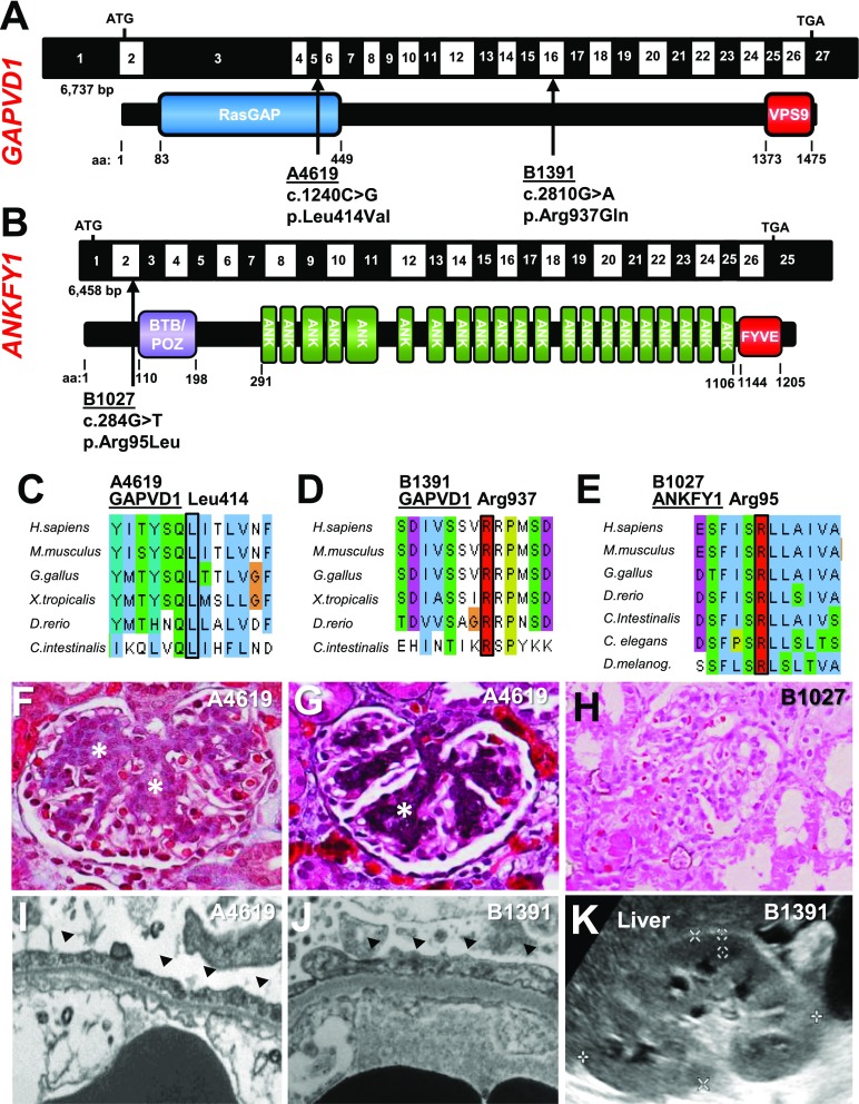
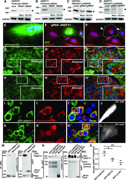
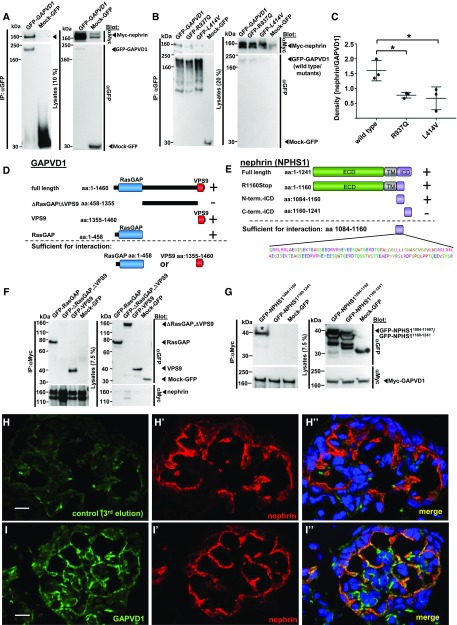
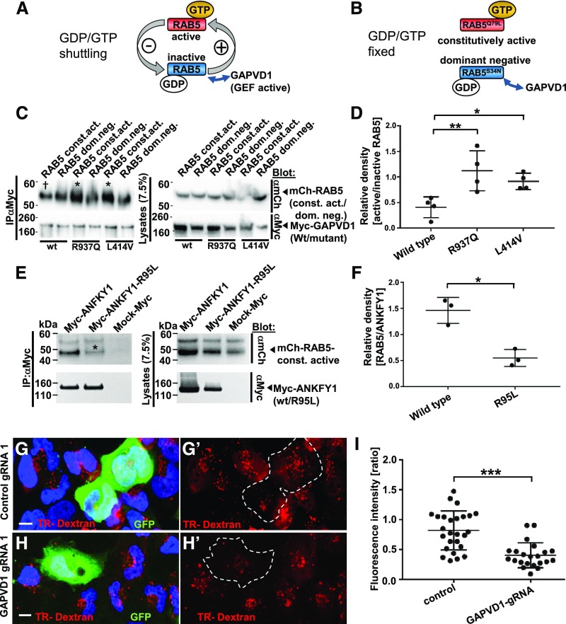
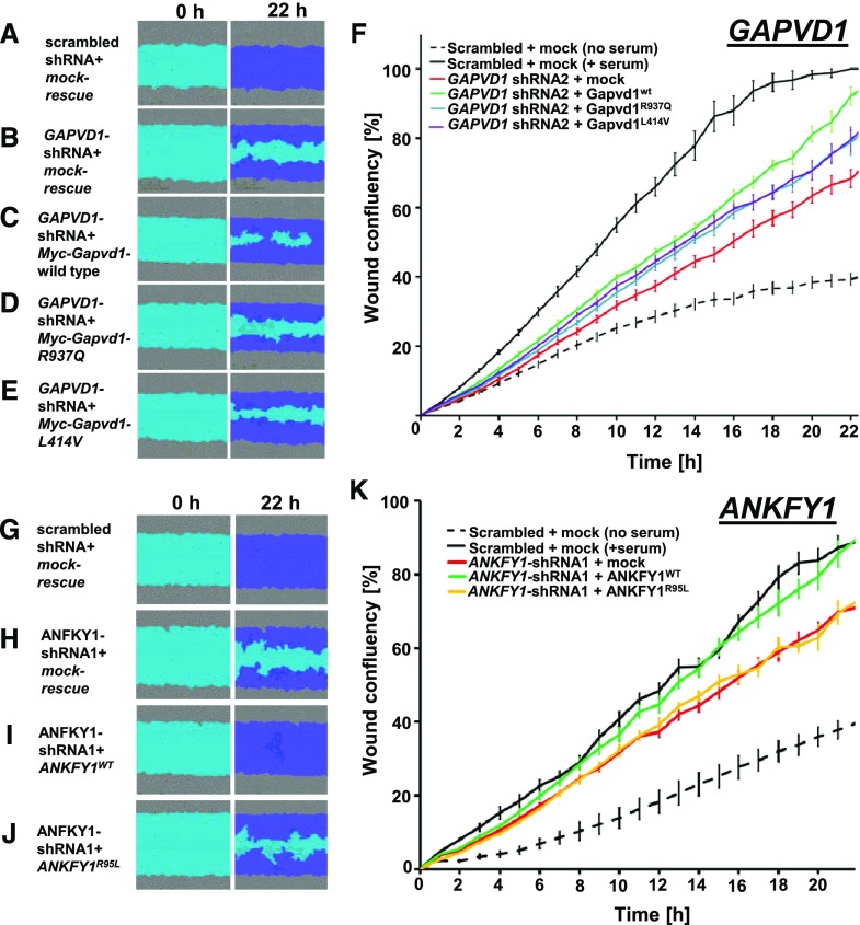
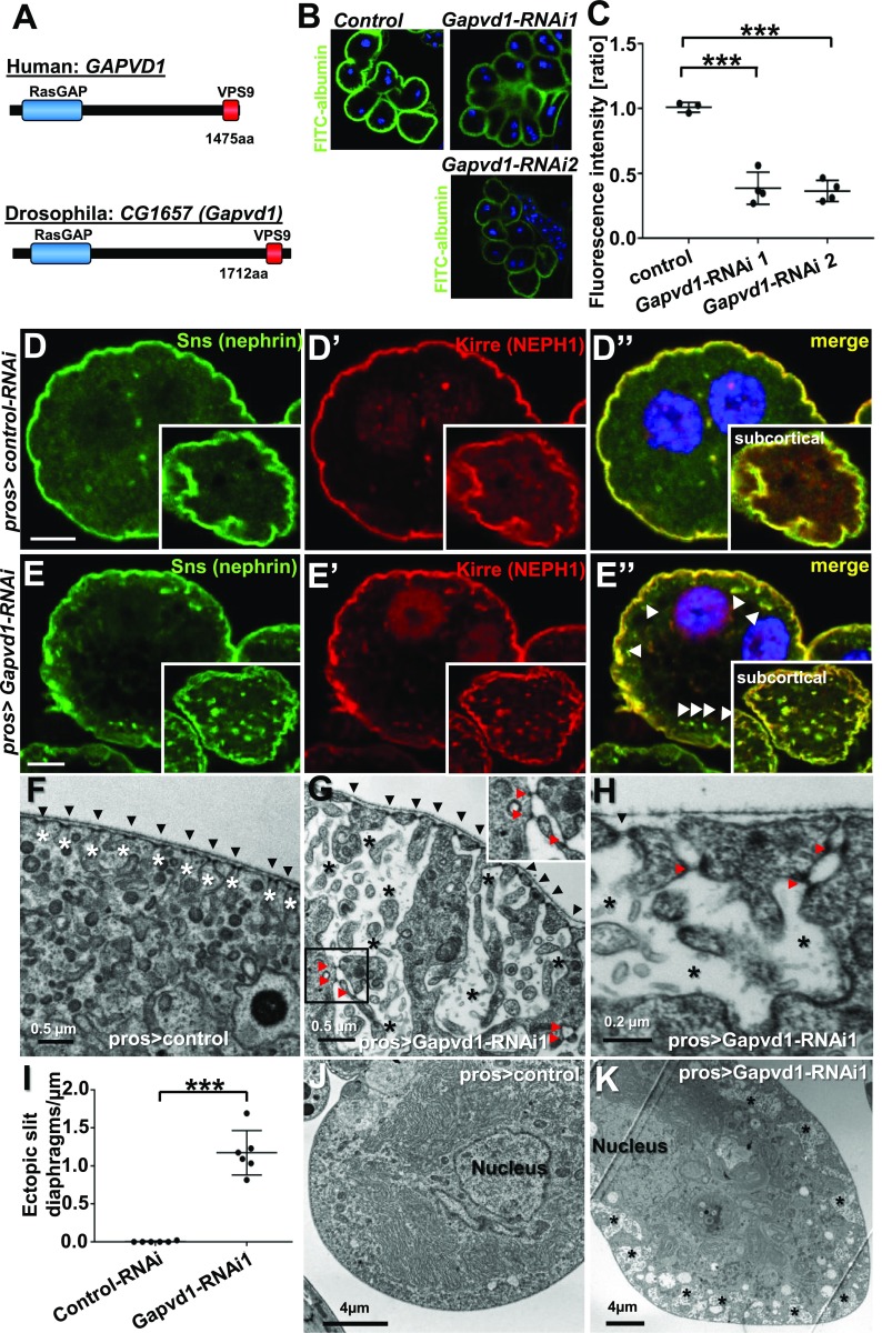
Results: We identified conserved, homozygous missense mutations of GAPVD1 in two families with early-onset NS and a homozygous missense mutation of ANKFY1 in two siblings with SRNS. GAPVD1 and ANKFY1 interact with the endosomal regulator RAB5. Coimmunoprecipitation assays indicated interaction between GAPVD1 and ANKFY1 proteins, which also colocalized when expressed in HEK293T cells. Silencing either protein diminished the podocyte migration rate. Compared with wild-type GAPVD1 and ANKFY1, the mutated proteins produced upon ectopic expression of GAPVD1 or ANKFY1 bearing the patient-derived mutations exhibited altered binding affinity for active RAB5 and reduced ability to rescue the knockout-induced defect in podocyte migration. Coimmunoprecipitation assays further demonstrated a physical interaction between nephrin and GAPVD1, and immunofluorescence revealed partial colocalization of these proteins in rat glomeruli. The patient-derived GAPVD1 mutations reduced nephrin-GAPVD1 binding affinity. In Drosophila, silencing Gapvd1 impaired endocytosis and caused mistrafficking of the nephrin ortholog.

Conclusions: Mutations in GAPVD1 and probably in ANKFY1 are novel monogenic causes of NS. The discovery of these genes implicates RAB5 regulation in the pathogenesis of human NS.

Keywords: endocytosis; genetic renal disease; nephrin; nephrocyte; nephrotic syndrome; podocyte.


Figures

None
Graphical abstract
Figure 1.
Figure 1.
WES identifies recessive disease-causing mutations in GAPVD1 and ANKFY1 in families with early-onset nephrotic syndrome. (A) Schematic of GAPVD1 cDNA with the corresponding protein including its functional domains. Arrows indicate the positions of two recessive mutations of GAPVD1 that were identified by WES in two families (B1391 and A4619) with nephrotic syndrome. (B) Schematic of ANKFY1 cDNA with the corresponding protein including its functional domains. Arrow indicates the position of one recessive missense mutation of ANKFY1 that was identified by WES in two individuals from family B1027 with nephrotic syndrome. (C) Alignment of GAPVD1 aa sequences for Homo sapiens, Mus musculus, Gallus gallus, Xenopus tropicalis, Danio rerio, and Ciona intestinalis shows conservation of the residue Leucine 414. (D) Alignment of aa sequence of GAPVD1 for H. sapiens, M. musculus, G. gallus, X. tropicalis, D. rerio, and C. intestinalis shows conservation of the residue Arginine 937. (E) Alignment of aa sequence of ANKFY1 for H. sapiens, M. musculus, G. gallus, X. tropicalis, D. rerio, C. intestinalis, C. elegans, and D. melanogaster shows conservation of the residue Arginine 95. (F) Renal histology (trichrome staining) of patient A4619 with the Leu414Val mutation of GAPVD1 shows mesangial hypercellularity and expansion of the extracellular matrix (asterisks). (G) Renal histology (Jones silver stain) of patient A4619 confirms extracellular matrix expansion (asterisk). (H) Renal histology of patient B1027 (HE staining) reveals FSGS. (I and J) Electron microscopy image shows podocyte foot process effacement (arrow heads) in (I) A4619 and (J) B1391. (K) Renal ultrasound image of B1391 shows increased echogenicity with renal echogenicity equal to liver echogenicity. aa, amino acid.
Figure 2.
Figure 2.
GAPVD1 and ANKFY1 proteins are expressed in a podocyte cell line and are interaction partners. (A) Transient, siRNAi-mediated silencing of GAPVD1 (arrow head) in human podocytes is demonstrated by immunoblot. This indicates endogenous expression of GAPVD1 in immortalized podocytes. (B) Transient, siRNAi-mediated silencing of ANKFY1 (arrow head) in human podocytes is demonstrated by immunoblot. This indicates endogenous expression of ANKFY1 in immortalized podocytes. (C) Western blot shows CRISPR/Cas9-mediated knockdown of GAPVD1 in HEK293T cells for two independent gRNAs (arrow head). This indicates endogenous expression of GAPVD1 in HEK293T cells and confirms the efficiency of the gRNAs. (D) Western blot shows CRISPR/Cas9-mediated silencing of ANKFY1 in HEK293T cells for two independent gRNAs (arrow head). This indicates endogenous expression of ANKFY1 in HEK293T cells and confirms the efficiency of the gRNAs. (E) Overexpressed N-GFP-GAPVD1 localizes to the cytosol in cultured podocytes. (F and F’) CRISPR/Cas9 mediated silencing of ANKFY1 in human podocytes is marked by coexpression of GFP (shown in [F]) and abrogates the signal from small, cytosolic vesicles using an anti-ANKFY1 antibody (gRNA-expressing cells are outlined by dashed line and marked by arrow heads in [F’]). (G and H) Staining human renal tissue sections with anti-ANKFY1 results in (G and G’’) a diffuse signal that was more intense than (H and H’’) the control signal (secondary antibody alone). Enhanced resolution microscopy by Airyscan technology revealed (insets G–G’’) fine vesicles in all glomerular cells including podocytes, whereas (H–H’’) no vesicular pattern was detected in the control. Nephrin staining is shown in (G’ and G’’) and (H’ and H’’). Scale bars represent 10 µm. (I and J) Overexpressed GFP-ANKFY1 and mCherry-GAPVD1 colocalize in HEK293T cells (I–I’’). (J) Colocalization of GFP-ANKFY1 and mCherry-GAPVD1 is confirmed by a scatter plot and a high Pearson’s coefficient (0.98). (K and L) Colocalization of GFP-ANKFY1 and an mCherry control appears incomplete (K–K’’). (L) Pearson coefficient is 0.35. (M) Upon overexpression and co-IP N-Myc–tagged GAPVD1 precipitates GFP-ANKFY1, but not Mock-GFP, in HEK293T cells. (N) Upon overexpression and co-IP with anti-GFP antibody, GFP-tagged GAPVD1 precipitates Myc-ANKFY1, but not Mock-GFP, in HEK293T cells. GAPVD1 cDNA constructs that reflect the mutations from patients B1391 (asterisk) and A4619 (dagger) show reduced binding affinity to Myc-ANKFY1. (O) Quantitation of density from (N) shows a significantly reduced affinity of mutant GAPVD1 (R937Q and L414V) to Myc-ANKFY1. Densitometry results from (N) were expressed as ANKFY1/GAPVD1wild type/mutant (n=3, P<0.05 for R937Q and P<0.01 for L414V). contr, control; IP, immunoprecipitation; PC, Pearson's coefficient; scra, scrambled; wt/mut, wild type/mutant.
Figure 3.
Figure 3.
The N-terminal cytosolic domain of nephrin interacts with the RasGAP and VPS9 domains of GAPVD1 and both proteins partially colocalize in neonatal rat kidney. (A) Upon overexpression in HEK293T cells and co-IP, GFP-tagged GAPVD1, but not Mock-GFP, precipitates Myc-tagged nephrin, yielding only the lower of two bands (arrow head). (B) GAPVD1 cDNA constructs that reflect the mutations from patients A4619 (p.Leu414Val) and B1391 (p.Arg937Gln) exhibit reduced binding affinity to nephrin. (C) Quantitation of density from (B) shows a significantly reduced affinity of GAPVD1 mutants to nephrin. Densitometry results from (B) were expressed as nephrin/GAPVD1wild type/mutant (n=3, P<0.05). (D) Schematic of truncation constructs of GAPVD1 and their ability to interact with nephrin (indicated by “+” versus “–,” also see below). (E) Schematic of truncation constructs of nephrin and their ability to interact with GAPVD1 (indicated by “+” versus “–,” also see below). (F) Upon overexpression and co-IP, full-length nephrin does not precipitate GFP-tagged GAPVD1 that lacks both functional domains (ΔRasGAP, ΔVPS9), whereas full-length nephrin precipitates the respective GAPVD1 constructs that contain the RasGAP or the VPS9 domain alone. This suggests that both functional domains of GAPVD1 show affinity for nephrin independently. (G) Mock-GFP and a truncated cDNA construct of nephrin (aa 1160–1241) that reflects the C-terminal half of the ICD of nephrin do not interact with GFP-GAPVD1. A GFP-tagged truncation construct (aa 1084–1160) that represents the N-terminal half of the ICD of nephrin interacts with Myc-tagged GAPVD1 (asterisk). This suggests that this subdomain of 76 aa mediates binding to GAPVD1. (H–I’’) Frozen sections of neonatal rat kidney were stained with anti-nephrin (red) and anti-GAPVD1 (green) or control. Nuclei are marked by Hoechst 33342 in blue. (I–I’’) Nephrin and GAPVD1 colocalize partially in newborn rat kidney. GAPVD1 is not restricted to podocytes but localizes to mesangial cells as well. (H–H’’) The third elution fraction obtained during the antibody purification of the GAPVD1 antibody (that contains only traces of antibody) was used as control and shows virtually no GAPVD1 signal. All images were recorded with identical confocal settings. Scale bars represent 10 µm. aa, amino acid; IP, immunoprecipitation; term., terminal.
Figure 4.
Figure 4.
Mutations of GAPVD1 that cause nephrotic syndrome increase the affinity to active RAB5, and GAPVD1 promotes dextran endocytosis. (A and B) Schematic showing that (A) RAB5 shuttles between active and inactive states dependent on binding to GTP/GDP, whereas (B) dominant negative and constitutively active RAB5 constructs are clamped to active and inactive states, respectively. (C) Overexpression and co-IP of mCherry-tagged RAB5 dominant negative (RAB5 dom. neg.) and constitutively active (RAB5 const. act.) constructs together with Myc-GAPVD1 reflecting the wild-type (WT) sequence or mutations causing nephrotic syndrome (R937Q and L414V). WT and mutant GAPVD1 interact with active and inactive RAB5. The mutant constructs of GAPVD1 show a stronger affinity to constitutively active RAB5 (asterisks) compared with WT GAPVD1 († sign). (D) Quantitation of density from precipitates analogous to (C) normalized to respectively precipitated RAB5 construct for co-IPs and shown as a ratio of RAB5 const. act. divided by RAB5 dom. neg. (n=4, P<0.05 or 0.01, respectively). (E) Overexpression and co-IP of mCherry-tagged constitutively active RAB5 together with Myc-ANFKY1 reflecting the WT sequence or the R95L mutation shows strongly reduced amounts of RAB5 precipitating with the mutant ANKFY1 (asterisk), indicating a reduced binding affinity. (F) Quantitation of density from precipitates of RAB5 protein analogous to (E) normalized to respectively precipitated Myc-ANKFY1 WT or mutant protein (n=3, P<0.05). (G–H’) Human podocytes transfected with plasmids expressing gRNA, Cas9, and GFP are exposed to Texas-Red-dextran (10 kD) for 30 minutes. Nuclei are marked by Hoechst 33342 in blue. (G and G’) Podocytes expressing control gRNA exhibit comparable Texas-Red-dextran endocytosis to the neighboring cells, whereas (H and H’) podocytes expressing a gRNA targeting GAPVD1 exhibit reduced tracer endocytosis. Scale bars represent 10 µm. (I) Quantitation of results from (G–H’). For both control gRNA or GAPVD1 targeting gRNA fluorescence intensity ratio is shown between gRNA-expressing cells and their nontransfected neighboring cells (n=2, approximately 25 cells each, P<0.001).
Figure 5.
Figure 5.
GAPVD1 and ANKFY1 regulate podocyte migration rate. (A–E) Podocyte migration rate is analyzed by IncuCyte videomicroscopy. Representative images show human podocytes after induction of scratch wound (light blue) and 22 hours thereafter. Scratch wound area (light blue) and podocytes that have migrated (dark blue) are shown at 22 hours. Serum addition strongly increases podocyte migration rate. Podocytes stably expressing scrambled shRNA (negative control) show (A) complete wound closure, whereas (B–F) silencing GAPVD1 results in reduced migration. The decrease in podocyte migration was (C) strongly reversed by transfection of mouse Gapvd1 but (D and E) only partially rescued by murine Gapvd1 constructs reflecting mutations R937Q and L414V detected in patients with nephrotic syndrome. (F) Graph shows wound confluence versus time for conditions described in (A–E). Error bars indicate SD of 12 wells with identical conditions (n=3). (G–J) Podocyte migration is observed, indicating (G) complete wound closure upon stable expression of scrambled shRNA (negative control), whereas (H) silencing ANKFY1 in the presence of a mock-rescue results in reduced migration. The decrease in podocyte migration was (I) strongly reversed by transfection of shRNA-resistant ANKFY1, whereas (J) introduction of the mutation from family B1027 (R95L) into the rescue construct abrogates this ability. (K) Graph shows wound confluence versus time for the conditions described in (G–J). Error bars indicate SD of 12 wells with identical conditions (n=3).
Figure 6.
Figure 6.
Silencing the Drosophila ortholog of GAPVD1 in nephrocytes affects slit diaphragm restriction and nephrocyte function. (A) Amino acid sequence of human GAPVD1 is 29% identical to the Drosophila ortholog CG1657 (Gapvd1), which shares the RasGAP and VPS9 domain as functional domains. (B) Silencing the GAPVD1 ortholog by two independent RNAi lines in nephrocytes using prospero-GAL4 significantly reduces uptake of FITC-albumin as an established assay of nephrocyte function. (C) Quantitation of data in (B) (n=3 per genotype, P<0.001). (D–D’’) Equatorial cross-section of a negative control garland cell nephrocyte costained for the nephrin ortholog Sns (green) and the KIRREL/NEPH1 ortholog Kirre (red). Slit diaphragm proteins localize at the cell periphery in a fine line. Inset shows a subcortical section; Sns and Kirre are restricted to the plasma membrane. Nuclei are marked by Hoechst 33342 in blue. Scale bar represents 5 µm. (E–E’’) Equatorial cross-section of garland cell nephrocytes expressing Gapvd1-RNAi shows appearance of vesicles (arrow heads) and broadening of the line of slit diaphragm proteins. Inset shows a subcortical section; Sns and Kirre are observed in puncta and are not restricted to the membrane. Nuclei are marked by Hoechst 33342 in blue. Scale bar represents 5 µm. (F) Nephrocyte expressing control-RNAi shows regular formation of slit diaphragms (black arrow heads). Labyrinthine channels are slender and restricted to the cortical area (white asterisks). (G and H) EM image from a section through the surface of a nephrocyte expressing Gapvd1-RNAi shows cross-section of slit diaphragms (black arrow heads) on the surface but also in an ectopic localization deeper in the labyrinthine channels (red arrow heads, see also inset). Labyrinthine channels are dilated, fused, and protrude deeply into the cell (white asterisks). (I) Quantitation of the number of ectopic intracellular slit diaphragms underneath the cell surface formed per micrometer of the cell surface length. Note that ectopic slit diaphragms are absent under negative control conditions, whereas more than one ectopic slit diaphragm per micrometer is formed upon expression of Gapvd1-RNAi 1 (quantitation of six cells from three different animals per genotype, P<0.001). (J and K) Compared with (J) control nephrocytes, (K) cells expressing Gapvd1-RNAi show cortical areas of lower electro-density (black asterisks). These areas correspond to the enlarged labyrinthine channels (see [G and H]). Pros, prospero-GAL4.

Similar articles

Cited by

References

    1. Bierzynska A, Soderquest K, Dean P, Colby E, Rollason R, Jones C, et al. .; NephroS; UK study of Nephrotic Syndrome : MAGI2 mutations cause congenital nephrotic syndrome. J Am Soc Nephrol 28: 1614–1621, 2017 - PMC - PubMed
    1. Lovric S, Goncalves S, Gee HY, Oskouian B, Srinivas H, Choi WI, et al. .: Mutations in sphingosine-1-phosphate lyase cause nephrosis with ichthyosis and adrenal insufficiency. J Clin Invest 127: 912–928, 2017 - PMC - PubMed
    1. Lovric S, Ashraf S, Tan W, Hildebrandt F: Genetic testing in steroid-resistant nephrotic syndrome: When and how? Nephrol Dial Transplant 31: 1802–1813, 2016 - PMC - PubMed
    1. Kestilä M, Lenkkeri U, Männikkö M, Lamerdin J, McCready P, Putaala H, et al. .: Positionally cloned gene for a novel glomerular protein--nephrin--is mutated in congenital nephrotic syndrome. Mol Cell 1: 575–582, 1998 - PubMed
    1. Löwik MM, Groenen PJ, Pronk I, Lilien MR, Goldschmeding R, Dijkman HB, et al. .: Focal segmental glomerulosclerosis in a patient homozygous for a CD2AP mutation. Kidney Int 72: 1198–1203, 2007 - PubMed

Publication types

MeSH terms

LinkOut - more resources