. 2018 Jun 29;9(7):734.
doi: 10.1038/s41419-018-0779-3.

The Hsp70 inhibitor 2-phenylethynesulfonamide inhibits replication and carcinogenicity of Epstein-Barr virus by inhibiting the molecular chaperone function of Hsp70

Affiliations

The Hsp70 inhibitor 2-phenylethynesulfonamide inhibits replication and carcinogenicity of Epstein-Barr virus by inhibiting the molecular chaperone function of Hsp70

Huan Wang et al. Cell Death Dis. .

Abstract

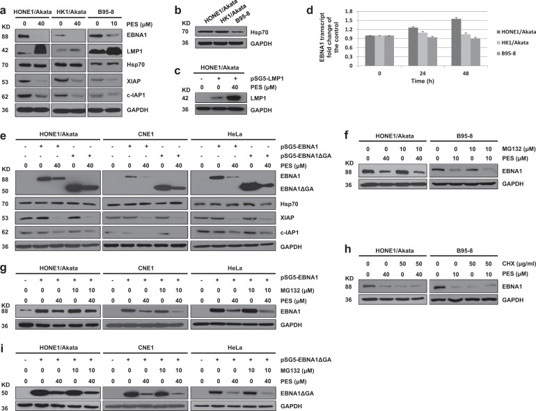
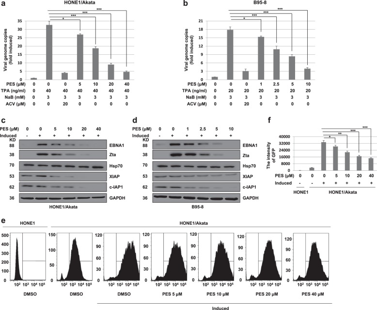
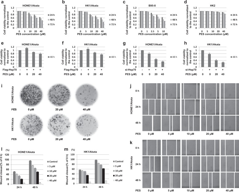
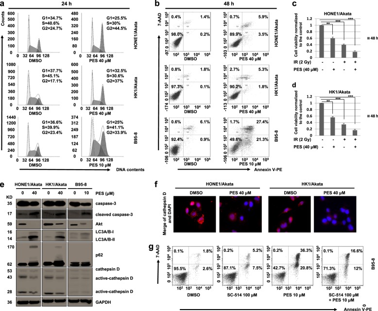
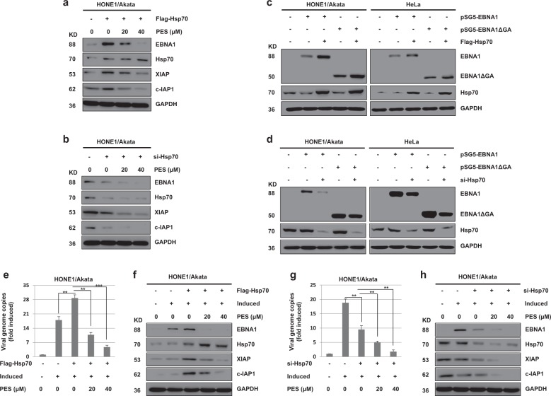
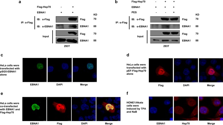
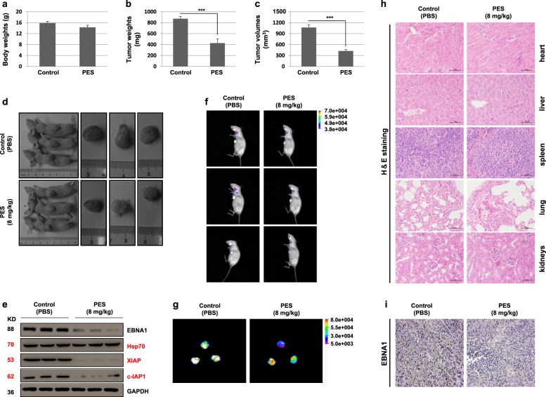
Epstein-Barr virus (EBV) can infect cells in latent and lytic period and cause serious disease. Epstein-Barr virus nuclear antigen 1 (EBNA1) is essential for the maintenance of the EBV DNA episome, replication and transcription. 2-phenylethynesulfonamide (PES) is a small molecular inhibitor of Heat shock protein 70 (Hsp70), which can interact with Hsp70 and disrupts its association with co-chaperones and substrate proteins of Hsp70. In our study, we found that PES could decrease the expression of EBNA1, which is independent of effects on EBNA1 transcription or proteasomal degradation pathway. The central glycine-alanine repeats domain was not required for inhibition of EBNA1 expression by PES. Also, PES could reduce the amount of intracellular EBV genomic DNA. PES inhibited proliferation and migration but induced cell cycle arrest and apoptosis of EBV positive cells. In addition, silencing of Hsp70 decreased expression of EBNA1 and the amounts of intracellular EBV genomic DNA, and PES increased this effect on a dose-dependent manner. On the contrast, over-expression of Hsp70 enhanced the expression of EBNA1 and the amounts of intracellular EBV genomic DNA, but PES inhibited this effect on a dose-dependent manner. Furthermore, Hsp70 interacted with EBNA1 but PES interfered this interaction. Our results indicate that PES suppresses replication and carcinogenicity of Epstein-Barr virus via inhibiting the molecular chaperone function of Hsp70.


Conflict of interest statement

The authors declare that they have no conflict of interest.

Figures

Fig. 1
Fig. 1. PES decreases EBNA1 expression and the GAr domain is not required for inhibition of EBNA1 expression by PES.
a HONE1/Akata, HK1/Akata, and B95-8 cells were treated with concentrations of PES as mentioned in Figure for 48 h. b HONE1/Akata, HK1/Akata, and B95-8 cells were untreated and examined the natural expression levels of Hsp70. c HONE1/Akata cells were transfected with empty vector pSG5or pSG5-LMP1, followed by a 44 h treatment with 40 µM PES beginning at 4 h after transfection. Whole-cell proteins were extracted and subjected to western blotting. d HONE1/Akata, HK1/Akata, and B95-8 cells were treated with drug vehicle control or PES (40, 40 or 10 μM) for 24 and 48 h, respectively. Total RNAs were extracted and reversely transcribed to cDNA. The level of EBNA1 transcript was examined by RT-PCR. The level of EBNA1 transcript in cells treated with vehicle control is set as 1. e HONE1/Akata, CNE1, and HeLa cells were transfected with empty vector, pSG5-EBNA1, or pSG5-EBNA1ΔGA for 4 h, followed by a 44 h treatment of 40 µM PES. f HONE1/Akata and B95-8 cells were treated with indicated concentrations of PES for 48 h in the absence or presence of proteasome inhibitor MG-132 (10 µM) for the last 16 h. g HONE1/Akata, CNE1, and HeLa cells were transfected with pSG5-EBNA1 for 4 h, followed by a 44 h treatment with 40 µM PES in the absence or presence of MG-132 (10 µM) at the last 16 h. h HONE1/Akata and B95-8 cells were treated with concentrations of PES as mentioned in the figure for 48 h in the absence or presence of CHX (50 μg/ml) at the last 12 h. i HONE1/Akata, CNE1, and HeLa cells were transfected with pSG5-EBNA1ΔGA, followed by a 48 h treatment with 40 µM PES beginning at 4 h after transfection in the absence or presence of MG-132 (10 µM) at the last 16 h. Whole-cell extracts were analyzed using western blotting
Fig. 2
Fig. 2. PES reduces lytic replication of EBV in HONE1/Akata and B95-8 cells.
a HONE1/Akata and b B95-8 cells were induced or not by TPA (40 or 20 ng/ml) and NaB (3 mM) for 4 h, followed with a 44 h treatment of ACV or gradient concentrations of PES. The intracellular genomic DNA were prepared and quantified by RT-PCR with EBNA1 primers. Each sample was normalized to the amount of the GAPDH gene. c HONE1/Akata and d B95-8 cells were induced by TPA and NaB for 4 h, and then treated with indicated concentrations of PES for 44 h. Whole-cell proteins were extracted and analyzed by western blotting. e,f HONE1/Akata cells were induced by TPA and NaB, followed by a 44 h treatment with increasing concentrations of PES beginning at 4 h after EBV induction. The untreated HONE1 cells and above-treated HONE1/Akata cells were collected and assessed by flow cytometer to analyze the intensity of GFP (*P < 0.05, **P < 0.01, ***P < 0.001)
Fig. 3
Fig. 3. PES inhibits proliferation and migration of EBV-positive cells.
a HONE1/Akata, b HK1/Akata, c B95-8 and d HK2 cells were treated or not with increasing concentrations of PES for 24, 48, and 72 h, respectively. e HONE1/Akata and f HK1/Akata cells were transfected with the pEF-Flag-Hsp70, followed by treatment with PES (20, 40 μM) beginning at 4 h after transfection. g HONE1/Akata and h HK1/Akata cells were transfected with Hsp70 siRNA, followed by treatment with PES (20, 40 μM) beginning at 4 h after transfection. Cell viability was determined by CCK-8 assays. i HONE1/Akata and HK1/Akata cells were treated or not with PES (20 or 40 μM) and cultured for another 15 days. Colony formation assay was carried out to determine the long-term effect of PES on cell proliferation. HONE1/Akata (j,l) and HK1/Akata (k,m) cells were treated with the linear scratch wounds, followed by the treatment of PES (0, 5, 10, 20 or 40 μM) for 24 and 48 h. Then the migration of the cells towards the wound was visualized. Photographs were captured and analyzed using Image J software
Fig. 4
Fig. 4. PES induces cell cycle arrest and apoptosis in EBV-positive cells.
a HONE1/Akata, HK1/Akata, and B95-8 cells were treated with indicated concentrations of PES for 24 h, followed by staining with PI. Cell cycle was assessed by flow cytometer. b HONE1/Akata, HK1/Akata and B95-8 cells were treated with concentrations of PES as mentioned above for 48 h and then stained with V-PE/7-AAD. Apoptotic cells analysis was performed using flow cytometer. c HONE1/Akata and d HK1/Akata cells were treated with 40 μM PES and then irradiated by a single dose of radiation (2 Gy). After 48 h, cell viability was determined by CCK-8 assays. e HONE1/Akata, HK1/Akata, and B95-8 cells were treated with concentrations of PES as mentioned above for 48 h. Whole-cell proteins were extracted and subjected to western blotting. f HONE1/Akata and HK1/Akata cells were treated with 40 μM PES for 48 h. Then cells were stained with cathepsin D antibody and visualized using the Leica confocal LCS-SP8-STED Nanoscope. g B95-8 cells were treated with SC-514 (100 μM) and PES (10 μM) for 48 h and then stained with V-PE/7-AAD. Apoptotic cells analysis was performed using flow cytometer (**P < 0.01, ***P < 0.001)
Fig. 5
Fig. 5
The change of EBNA1 expression induced by Hsp70 is not associated with the GAr domain, and Hsp70 promote the lytic replication of EBV in HONE1/Akata cells but PES inhibits this effect. HONE1/Akata cells were transfected with pEF-Flag-Hsp70 (a) or Hsp70 siRNA (b) for 24 h, followed by treatment with PES (20 or 40 μM) for 48 h. c HONE1/Akata and HeLa cells were transfected with pEF-Flag-Hsp70 for 24 h, followed by transfection with pSG5-EBNA1 or pSG5-EBNA1ΔGA into above cells for 48 h. d HONE1/Akata and HeLa cells were transfected with Hsp70 siRNA for 24 h, followed by transfection with pSG5-EBNA1 or pSG5-EBNA1ΔGA into above cells for 48 h. Whole-cell proteins above were extracted and subjected to western blotting. HONE1/Akata cells were induced by TPA and NaB for 4 h, and then transfected with pEF-Flag-Hsp70 (e) or Hsp70 siRNA (g), followed by treatment with PES beginning at 4 h after transfection. The intracellular genomic DNA were quantified by RT-PCR with EBNA1 primers. Each sample was normalized to the amount of the GAPDH gene. f,h Whole-cell proteins above were extracted and analyzed using western blotting (**P < 0.01, ***P < 0.001)
Fig. 6
Fig. 6. EBNA1 interacts with Hsp70 but PES interferes this interaction.
a 293T cells were transfected with pEF-Flag-Hsp70 in the presence of pSG5-EBNA1. At 48 h post-transfection, IP assays were performed with Flag antibody, and then immunoprecipitated with the indicated antibodies. b 293T cells were transfected with pSG5-EBNA1 and pEF-Flag-Hsp70, separately or together, followed by treatment with the vehicle drug control or PES (20 μM) beginning at 4 h after transfection. 48 h later, IP assays were performed. HeLa cells were transfected with pSG5-EBNA1 (c) and pEF-Flag-Hsp70 alone (d) and co-transfected with pSG5-EBNA1 and pEF-Flag-Hsp70 (e). f HONE1/Akata cells were induced by TPA and NaB. At 48 h post-transfection or post-induction, cells were fixed, stained with antibodies, and then visualized using the Leica confocal LCS-SP8-STED Nanoscope
Fig. 7
Fig. 7. PES inhibits the growth of tumor in vivo.
BALB/c nude mice with hypodermic tumors were treated with vehicle control (PBS) or 8 mg/kg PES for 5 days. Body weights (a), tumor weights (b), and tumor volumes (c) were evaluated in above BALB/c nude mice. The whole values are shown as mean ± SD of six mice. d Representative images of mice treated with PBS or PES and tumors in above mice are shown. e Lysates of tumor tissues were analyzed by western blotting. f, g The intensity of GFP in tumors of above mice were assessed by In-vivo Xtreme Imaging System. h The five important organs (heart, liver, spleen, lung, and kidneys) of above mice were detected by H&E staining. i The tumor tissues were detected by immunohistochemical staining using antibody against EBNA1. Randomly selected micrographs were given (***P < 0.001)

References

    1. Wiedmer A, et al. Epstein–Barr virus immediate-early protein Zta co-opts mitochondrial single-stranded DNA binding protein to promote viral and inhibit mitochondrial DNA replication. J. Virol. 2008;82:4647–4655. doi: 10.1128/JVI.02198-07. - DOI - PMC - PubMed
    1. Sun X, et al. Hsp90 inhibitors block outgrowth of EBV-infected malignant cells in vitro and in vivo through an EBNA1-dependent mechanism. Proc. Natl. Acad. Sci. USA. 2010;107:3146–3151. doi: 10.1073/pnas.0910717107. - DOI - PMC - PubMed
    1. Whitehurst, C. B. et al. Knockout of Epstein–Barr virus BPLF1 retards B-cell transformation and lymphoma formation in humanized mice. mBio. 6, e01574-01515 (2015). - PMC - PubMed
    1. Raab-Traub N. Epstein–Barr virus and nasopharyngeal carcinoma. Semin. Cancer Biol. 1992;3:297–307. - PubMed
    1. Wang FZ, et al. Maribavir inhibits Epstein–Barr virus transcription in addition to viral DNA replication. J. Virol. 2009;83:12108–12117. doi: 10.1128/JVI.01575-09. - DOI - PMC - PubMed

Publication types

MeSH terms

Substances

LinkOut - more resources