. 2018 Jun 29;8(1):9822.
doi: 10.1038/s41598-018-28091-9.

Differential preservation of endogenous human and microbial DNA in dental calculus and dentin

Affiliations

Differential preservation of endogenous human and microbial DNA in dental calculus and dentin

Allison E Mann et al. Sci Rep. .

Abstract

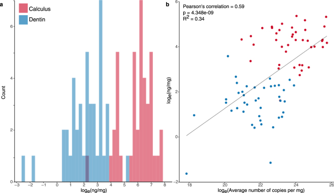
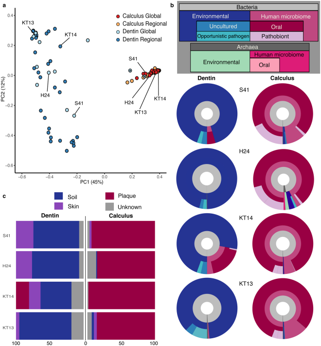
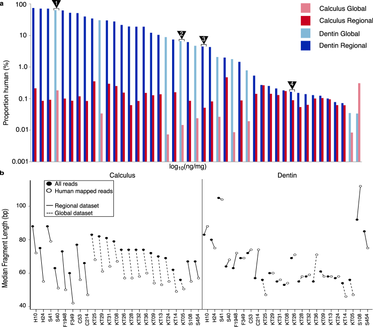
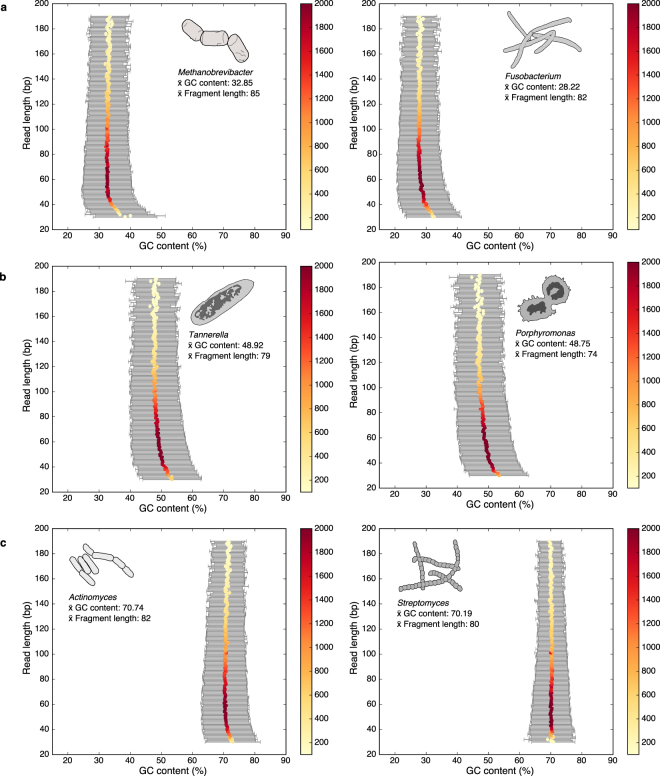
Dental calculus (calcified dental plaque) is prevalent in archaeological skeletal collections and is a rich source of oral microbiome and host-derived ancient biomolecules. Recently, it has been proposed that dental calculus may provide a more robust environment for DNA preservation than other skeletal remains, but this has not been systematically tested. In this study, shotgun-sequenced data from paired dental calculus and dentin samples from 48 globally distributed individuals are compared using a metagenomic approach. Overall, we find DNA from dental calculus is consistently more abundant and less contaminated than DNA from dentin. The majority of DNA in dental calculus is microbial and originates from the oral microbiome; however, a small but consistent proportion of DNA (mean 0.08 ± 0.08%, range 0.007-0.47%) derives from the host genome. Host DNA content within dentin is variable (mean 13.70 ± 18.62%, range 0.003-70.14%), and for a subset of dentin samples (15.21%), oral bacteria contribute > 20% of total DNA. Human DNA in dental calculus is highly fragmented, and is consistently shorter than both microbial DNA in dental calculus and human DNA in paired dentin samples. Finally, we find that microbial DNA fragmentation patterns are associated with guanine-cytosine (GC) content, but not aspects of cellular structure.


Conflict of interest statement

The authors declare no competing interests.

Figures

Figure 1
Figure 1
Geographic locations and temporal periods of archaeological teeth included in this study. (a) Camino del Molino, Iberia; (b) Arbulag Soum, Khövsgöl, Mongolia; (c) Samdzong, Nepal; (d) Kilteasheen, Ireland; (e) Anse à la Gourde, Guadeloupe; (f) Norris Farms, Illinois, USA; (g) Middenbeemster, the Netherlands. For each site, representative teeth with dental calculus deposits are shown in boxes. The number of teeth (dentin-calculus pairs) analyzed per site is provided within the indicated circles, and corresponding letters on the timeline indicate the time period represented by each site.
Figure 2
Figure 2
Total DNA content of dental calculus is higher than dentin as measured by both fluorescence and quantitative PCR (qPCR) techniques. (a) Normalized DNA yield (log transformed nanograms DNA per milligram starting material) of DNA extracts obtained from dental calculus and dentin as measured by a Qubit fluorometer using a High Sensitivity Assay (p = 3.911e-11, Wilcoxon signed-rank paired test). (b) Linear correlation between normalized DNA yield (log(ng/mg)) and normalized DNA yield as measured by qPCR after library preparation. The average number of copies per milligram was calculated from the mean of four technical replicates, and these values were compared between dentin and calculus (p = 6.941e-07, Wilcoxon signed-rank paired test). In both quantification metrics dental calculus has a higher DNA yield than dentin.
Figure 3
Figure 3
Microbial communities represented in archaeological dental calculus and dentin are distinct. (a) Principal Coordinates Analysis (PCoA) of Bray-Curtis distances of all bacterial and archaeal species-level assignments from dental calculus and dentin. Color indicates material type (dental calculus or dentin) and membership in global or regional dataset. Microbial taxonomy was assigned using MALT with the full NCBI nucleotide database. Dentin samples marked with an asterisk belong to individuals NF47 and NF217 in the global dataset, and may represent teeth with carious lesions (Supplementary Figure S2). (b) A subset of four sample pairs from the global (S41, H24) and regional (KT14, KT13) sample sets were selected to further demonstrate differences in dental calculus and dentin microbial communities. Species represented in the MALT results were sorted into a layered classification scheme (see legend in gray box), and the proportions of reads assigned to each taxon were used to generate Krona plots. (c) Stacked bar plots of Bayesian SourceTracker results for the four selected pairs display estimated proportions of source contribution at the genus level, using modern plaque, skin, and soil datasets as model sources. Both approaches show overwhelming abundance of environmental bacteria within dentin samples, while most microbial DNA within the paired calculus samples are associated with, and most likely derive from, the human oral microbiome. The robustness of the human microbiome signal to the environmental signal is driving the separation we see in the PCoA.
Figure 4
Figure 4
Human DNA in dental calculus shows consistent patterns of low relative abundance and high fragmentation. (a) Relative percentage of human DNA in all paired calculus and dentin samples calculated from de-duplicated reads mapped to the hg19 human reference genome using BWA. While the majority of dentin samples have an overall higher percentage of human DNA, this value varies substantially by sample. Calculus is comparatively consistent between samples albeit on average lower than their paired dentin sample. Sample pairs corresponding to those in Fig. 3 are indicated by numbered triangles: (1) S41, (2) H24, (3) KT14, (4) KT13. (b) Median fragment length of merged reads mapping to the human genome compared to all merged reads in both dental calculus and dentin. Human assigned reads in dental calculus are shorter than expected independent of age, laboratory processing protocol, or sample preservation. Human mapped reads in dental calculus and dentin were further verified for authenticity using strict mapping parameters (Supplementary Figure S5, Supplementary Table S9).
Figure 5
Figure 5
Fragment length and damage rates among bacterial taxa within calculus. (a) Fragment length distribution of 50 high frequency species-level bacteria among all dental calculus grouped into three metadata categories: gram status, the presence or absence of a surface layer (S-layer), and the overall genomic GC content of the organism as documented from the reference genome in the NCBI database. Input was normalized to 400 randomly chosen reads per sample per species to mitigate the impact of sample specific read length profiles. No detectable differences in the median are apparent for the presence of an S-layer or gram status. Genomic GC content, however, does impact the overall fragment length profile of reads. (b) Deviation of the median fragment length from overall sample median fragment length of a subset of 20 oral bacteria in dental calculus colored by dataset origin. Again, genomic GC content appears to impact the fragment length profiles of individual taxa where high GC content species have shorter overall fragment length profiles. (c) Terminal cytosine damage rates (C to T substitution ratio at the first position of the 5′ end of the molecule) among 50 bacterial species in dental calculus grouped by gram status, presence or absence of an S-layer, and overall genomic GC content. No detectable differences in damage rates could be detected in any of the three chosen metadata categories. (d) Terminal cytosine damage rates (C to T substitution ratio at the first position of the 5′ end of the molecule) among 20 oral bacteria. While some differences in damage patterns can be detected, it is unclear if this is random or due to some process not examined in this study.
Figure 6
Figure 6
Relationship of GC content to fragment length in five prevalent oral genera and one soil genus (Streptomyces). (a) Two low expected GC content genera, Methanobrevibacter and Fusobacterium binned by read lengths wherein each dot represents the mean GC content for that bin. (b) Two moderate expected GC content general, Tannerella and Porphyromonas binned by read lengths wherein each dot represents the mean GC content for that bin. (c) One high expected GC content oral genus (Actinomyces) and one high expected GC content soil genus (Streptomyces) binned by read lengths wherein each dot represents the mean GC content for that bin. Heat bar indicated the number of reads representing each length bin. In all cases except for Streptomyces only reads from dental calculus samples were included. Reads for Streptomyces were extracted from dentin samples to contrast GC shifts in a potentially modern soil contaminate with typical members of the human oral microbiome. In all cases except for Streptomyces the GC content of shorter reads is skewed higher than the overall mean value. This effect greater in low to moderate expected GC content genera than those with higher GC content (Supplementary Figure S8).

References

    1. White DJ. Processes contributing to the formation of dental calculus. Biofouling. 1991;4:209–218. doi: 10.1080/08927019109378211. - DOI
    1. Marsh, P. D. Dental plaque as a biofilm and microbial community - implications for health and disease. BMC Oral Health6, 10.1186/1472-6831-6-S1-S14 (2006). - PMC - PubMed
    1. Warinner C, Speller C, Collins MJ. A new era in palaeomicrobiology: prospects for ancient dental calculus as a long-term record of the human oral microbiome. Phil. Trans. R. Soc. B. 2015;370:20130376. doi: 10.1098/rstb.2013.0376. - DOI - PMC - PubMed
    1. Marsh PD, Bradshaw DJ. Dental plaque as a biofilm. Journal of Industrial Microbiology. 1995;15:169–175. doi: 10.1007/BF01569822. - DOI - PubMed
    1. Power RC, Salazar-García DC, Wittig RM, Freiberger M, Henry AG. Dental calculus evidence of plant diet and life history transitions in the chimpanzees of the Taï Forest. Scientific Reports. 2015;5:15161. doi: 10.1038/srep15161. - DOI - PMC - PubMed

Publication types

LinkOut - more resources