

miR-153 inhibits the migration and the tube formation of endothelial cells by blocking the paracrine of angiopoietin 1 in breast cancer cells
- PMID: 29959560
- PMCID: PMC6208884
- DOI: 10.1007/s10456-018-9630-9
miR-153 inhibits the migration and the tube formation of endothelial cells by blocking the paracrine of angiopoietin 1 in breast cancer cells
Abstract
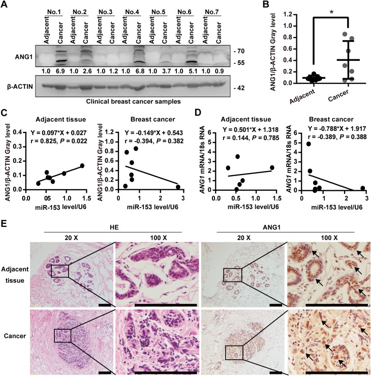
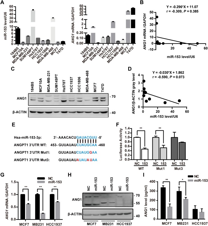
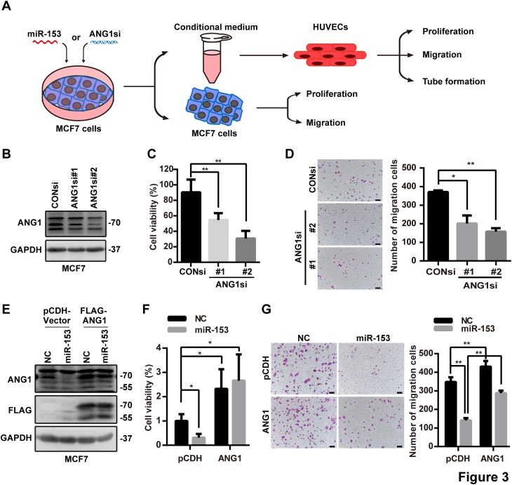
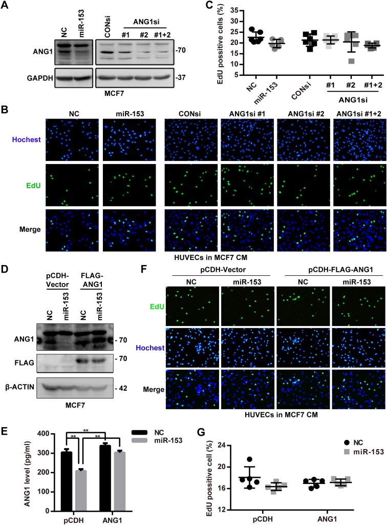
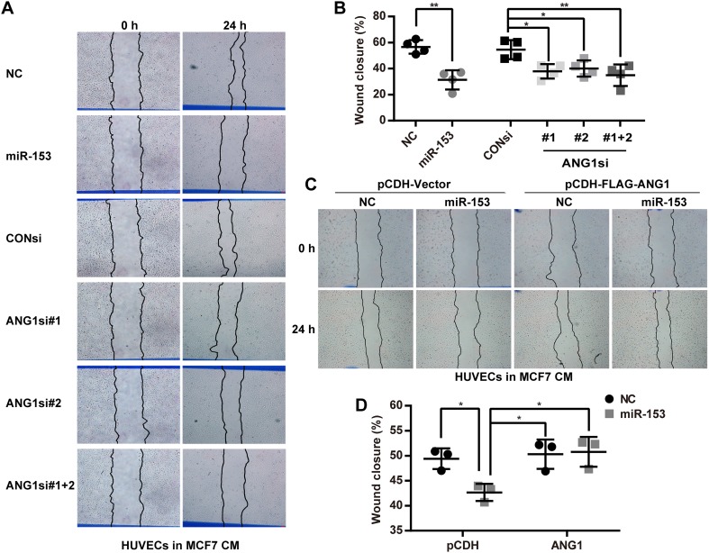
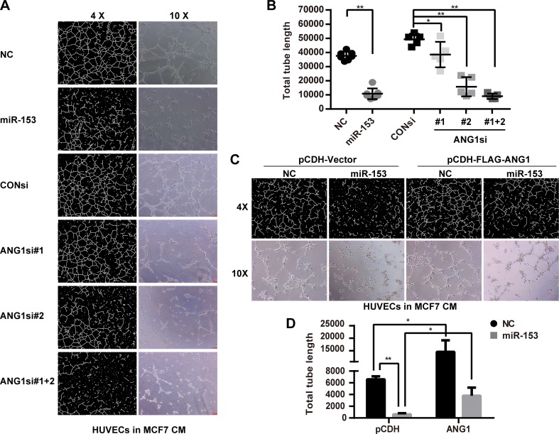
The sprouting of endothelial cells is the first step of tumor angiogenesis. Our previous study suggests that miR-153 suppresses breast tumor angiogenesis partially through targeting hypoxia-induced factor (HIF1α). In this study, we demonstrated that miR-153 also suppresses the migration and the tube formation of endothelial cells through directly targeting angiopoietin 1 (ANG1) in breast cancer cells. There was a negative correlation between miR-153 and ANG1 levels in breast cancer. miR-153 blocked the expression and secretion of ANG1 in breast cancer cells through binding to ANG1 mRNA. Conditioned medium from the breast cancer cell, MCF7, treated with miR-153 had no effect on the proliferation of HUVECs, but significantly inhibited the migration and tube formation of HUVECs, which could be rescued by overexpression of ANG1. In addition, miR-153 also directly inhibited the proliferation and migration of MCF7 through downregulation of ANG1. These findings suggest that miR-153 suppresses the activity of tumor cells and the migration and tube formation of endothelial cells by silencing ANG1.
Keywords: Angiopoietin 1; Breast cancer; Endothelial cell; MiR-153; Tumor angiogenesis.
Conflict of interest statement
The authors declare that no competing interest exists.
Figures
Similar articles
-
Dual targeting of ANGPT1 and TGFBR2 genes by miR-204 controls angiogenesis in breast cancer.Sci Rep. 2016 Oct 5;6:34504. doi: 10.1038/srep34504. Sci Rep. 2016. PMID: 27703260 Free PMC article.
-
Annexin-A1 regulates microRNA-26b* and microRNA-562 to directly target NF-κB and angiogenesis in breast cancer cells.PLoS One. 2014 Dec 23;9(12):e114507. doi: 10.1371/journal.pone.0114507. eCollection 2014. PLoS One. 2014. PMID: 25536365 Free PMC article.
-
MicroRNA-335 suppresses the proliferation, migration, and invasion of breast cancer cells by targeting EphA4.Mol Cell Biochem. 2018 Feb;439(1-2):95-104. doi: 10.1007/s11010-017-3139-1. Epub 2017 Aug 9. Mol Cell Biochem. 2018. PMID: 28795314
-
Role of miRNAs in tumor and endothelial cell interactions during tumor progression.Semin Cancer Biol. 2020 Feb;60:214-224. doi: 10.1016/j.semcancer.2019.07.024. Epub 2019 Aug 3. Semin Cancer Biol. 2020. PMID: 31386907 Review.
-
Role of miR-155 in breast cancer.Front Biosci (Landmark Ed). 2012 Jun 1;17(6):2350-5. doi: 10.2741/4056. Front Biosci (Landmark Ed). 2012. PMID: 22652783 Review.
Cited by
-
Targeting Angiogenesis in Breast Cancer: Current Evidence and Future Perspectives of Novel Anti-Angiogenic Approaches.Front Pharmacol. 2022 Feb 25;13:838133. doi: 10.3389/fphar.2022.838133. eCollection 2022. Front Pharmacol. 2022. PMID: 35281942 Free PMC article. Review.
-
MicroRNA-934 facilitates cell proliferation, migration, invasion and angiogenesis in colorectal cancer by targeting B-cell translocation gene 2.Bioengineered. 2021 Dec;12(2):9507-9519. doi: 10.1080/21655979.2021.1996505. Bioengineered. 2021. PMID: 34699325 Free PMC article.
-
A novel tubulin inhibitor, 6h, suppresses tumor-associated angiogenesis and shows potent antitumor activity against non-small cell lung cancers.J Biol Chem. 2022 Jul;298(7):102063. doi: 10.1016/j.jbc.2022.102063. Epub 2022 May 23. J Biol Chem. 2022. PMID: 35618020 Free PMC article.
-
MicroRNA‑218 inhibits tumor angiogenesis of human renal cell carcinoma by targeting GAB2.Oncol Rep. 2020 Nov;44(5):1961-1970. doi: 10.3892/or.2020.7759. Epub 2020 Sep 8. Oncol Rep. 2020. PMID: 32901879 Free PMC article.
-
Lysyl oxidase and hypoxia-inducible factor 1α: biomarkers of gastric cancer.World J Gastroenterol. 2019 Apr 21;25(15):1828-1839. doi: 10.3748/wjg.v25.i15.1828. World J Gastroenterol. 2019. PMID: 31057297 Free PMC article.
References
-
- Warren BA, Shubik P. The growth of the blood supply to melanoma transplants in the hamster cheek pouch. Lab Investig. 1966;15(2):464–478. - PubMed
-
- Folkman J. How is blood vessel growth regulated in normal and neoplastic tissue? G.H.A. Clowes memorial Award lecture. Cancer Res. 1986;46(2):467–473. - PubMed
Publication types
MeSH terms
Substances
LinkOut - more resources
Full Text Sources
Other Literature Sources
Medical
Miscellaneous
