. 2018 Sep;144(9):1685-1699.
doi: 10.1007/s00432-018-2696-3. Epub 2018 Jun 29.

Disruption of MEK/ERK/c-Myc signaling radiosensitizes prostate cancer cells in vitro and in vivo

Affiliations

Disruption of MEK/ERK/c-Myc signaling radiosensitizes prostate cancer cells in vitro and in vivo

Carmela Ciccarelli et al. J Cancer Res Clin Oncol. 2018 Sep.

Abstract

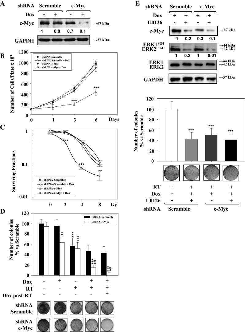
Purpose: Prostate cancer (PCa) cell radioresistance causes the failure of radiation therapy (RT) in localized or locally advanced disease. The aberrant accumulation of c-Myc oncoprotein, known to promote PCa onset and progression, may be due to the control of gene transcription and/or MEK/ERK-regulated protein stabilization. Here, we investigated the role of MEK/ERK signaling in PCa.

Methods: LnCAP, 22Rv1, DU145, and PC3 PCa cell lines were used in in vitro and in vivo experiments. U0126, trametinib MEK/ERK inhibitors, and c-Myc shRNAs were used. Radiation was delivered using an x-6 MV photon linear accelerator. U0126 in vivo activity alone or in combination with irradiation was determined in murine xenografts.

Results: Inhibition of MEK/ERK signaling down-regulated c-Myc protein in PCa cell lines to varying extents by affecting expression of RNA and protein, which in turn determined radiosensitization in in vitro and in vivo xenograft models of PCa cells. The crucial role played by c-Myc in the MEK/ERK pathways was demonstrated in 22Rv1 cells by the silencing of c-Myc by means of short hairpin mRNA, which yielded effects resembling the targeting of MEK/ERK signaling. The clinically approved compound trametinib used in vitro yielded the same effects as U0126 on growth and C-Myc expression. Notably, U0126 and trametinib induced a drastic down-regulation of BMX, which is known to prevent apoptosis in cancer cells.

Conclusions: The results of our study suggest that signal transduction-based therapy can, by disrupting the MEK/ERK/c-Myc axis, reduce human PCa radioresistance caused by increased c-Myc expression in vivo and in vitro and restores apoptosis signals.

Keywords: C-Myc; ERKs; Prostate cancer; Radioresistance; Radiotherapy; U0126.


Conflict of interest statement

The authors have no affiliations to disclose, including any of a financial nature, that they consider to be relevant or related to the subject matter discussed.

Figures

Fig. 1
Fig. 1
c-Myc and upstream c-Myc kinases expression in normal prostate and PCa cell lines. a Levels of c-Myc mRNA assessed by qRT-PCR in the various PCa cell lines expressed as fold changes vs. RNA level of normal RWPE1 taken as 1. Results (mean + SEM, Student’s t test, *P < 0.05; **P < 0.01; ***P < 0.001) are derived from three different low passages (from 5 to 8) for each cell line. b Upper panel, evaluation of c-Myc, phospho-ERKs, phospho-GSK3-beta protein levels in PCa cell lines vs. RWPE1 normal cells after western blot (lower panel). Antibodies to ERKs, GSK3, and GAPDH were used for normalization
Fig. 2
Fig. 2
Time course of c-Myc mRNA and protein in untreated and U0126-treated PCa cell lines. a c-Myc RNA was evaluated by qRT-PCR after different treatment periods. Each value is expressed as fold change vs. 2 h taken as 1. Results (mean ± SEM, Student’s t test, *P < 0.05; **P < 0.01; ***P < 0.001) are from a representative experiment of three. b PCa cell lines were treated with U0126 for different time periods. Phospho-ERKs and c-Myc values were calculated by measuring band intensity after immunoblotting using Image-J and normalizing phospho-ERKs with total ERKs and GAPDH and C-Myc with GAPDH. The same filter was used first for phospho-ERK and ERK immunodetection, with monoclonal and polyclonal antibody, respectively, and after stripping for C-Myc and GAPDH (see “Materials and methods”)
Fig. 3
Fig. 3
Effects of U0126 on PCa cell line radioresistance. a Cells were treated with RT, radiation alone; U-RT, 1-h pre-treatment with U0126 followed by radiation; U-RT-U, 1-h pre-treatment with U0126, followed by radiation, then by persistent U0126 treatment. Radiation doses ranged from 2 to 8 Gy. Cells were treated after plating in clonogenic conditions and stopped after 15 days of culture. The surviving fraction was calculated as colonies counted/cells seeded (plating efficiency). Results (mean ± SEM) are expressed as fold increase vs. RT-treated cells, taken as 1 (Student’s t test, *P < 0.05; **P < 0.01; ***P < 0.001). These are representative results of the effects of different RT doses combined or not combined with U0126 treatment on PCa cell lines in three different experiments
Fig. 4
Fig. 4
Effects of c-Myc silencing on growth, clonogenic assay, and radiation response of 22Rv1. a 22Rv1 expressing scramble or c-Myc shRNA under the control of doxycycline was established and cultured with or without doxycycline to measure c-Myc protein expression by western blot. Results of C-Myc level changes are expressed as fold increase vs. shRNA-scramble transfected cells, taken as 1. b Growth potential of scramble or c-Myc shRNA cells in the presence or absence of doxycycline. Each point is the result of three plates (mean ± SEM, Student’s t test, *P < 0.05; **P < 0.01, ***P < 0.001). Two different experiments were performed. c Scramble or c-Myc shRNA expressing cells were cultured and irradiated (2–8 Gy), doxycycline was added before radiation, the surviving fraction was calculated as colonies counted/cells seeded (plating efficiency). Results (mean + SEM) are derived from three experiments and expressed as fold increase vs. shRNA-scramble transfected cells, taken as 1 (Student’s t test, *P < 0.05; **P < 0.01; ***P < 0.001). d Clonogenic assay of c-Myc shRNA silenced or shRNA-scramble infected cells treated or not treated with doxycyclin and RT. Upper panel, histogram of colonies from different culture conditions. Colonies with more than 50 cells were counted and plotted. Results (mean ± SEM) are derived from three plates and expressed as fold increase vs. shRNA-scramble untreated (Dox-RT-Dox post-RT-) (Student’s t test, *P < 0.05; **P < 0.01, ***P < 0.001), vs. Dox-RT + Dox post-RT- (Student’s t test, $P < 0.05; $$P < 0.01, $$$P < 0.001) vs. Dox + RT + Dox postRT- (Student’s t test, #P < 0.05; ##P < 0.01, ###P < 0.001); lower panel, picture of plates with clones by scramble or c-Myc-shRNA cultured in the absence or in the presence of doxycycline (Dox), after 4 Gy radiation alone (RT), after pre-treatment of doxycycline and 4 Gy radiation (Dox-RT), or after pre-treatment of doxycycline followed first by 4 Gy radiation, then by persistent doxycycline treatment (Dox-RT-Dox post-RT)
Fig. 5
Fig. 5
MEK inhibitor reduces tumor growth of 22Rv1 cells transplanted mice. a Tumor from mice transplanted with parental 22Rv1 cells (UNTREATED), tumor from RT-treated mice (RT), tumor from U0126-treated mice (U0126), tumor from U0126-pre-treated and irradiated mice (U0126 + RT). U0126 treatment was stopped 22 days before the end point. Results (mean ± SEM) are expressed as fold increase vs. UNTREATED (Student’s t test, *P < 0.05; **P < 0.01; ***P < 0.001), vs. RT (Student’s t test, $P < 0.05; $$P < 0.01; $$$P < 0.001), vs. U0126 (Student’s t test, #P < 0.05; ##P < 0.01; ###P < 0.001). b Tumor weight of group of mice that were not treated (UNT) or were treated with U0126 (U0126), radiotherapy (RT) or with the combined treatment.(U0126 + RT). c Percentage of mice with tumor progression for UNTREATED, RT, U0126, and the combined treatment in 22Rv1 tumors. d Kaplan–Meier estimates for rates of progression in untreated, RT treated, U0126 treated and the combined treatment in 22Rv1 tumors. e Left panel, biochemical evidence of c-Myc and phospho-ERK expression in tumors from U0126-treated mice (U1, U2) and from RT-treated mice (RT1, RT2) or U0126 and irradiated mice (U + RT1, U + RT2). c-Myc values were normalized as c-Myc/GAPDH; the ERK-PO4 were normalized as ERKs-PO4/ERKs/GAPDH. The values of untreated cells were taken as 1. Right panel is a representative evaluation of the western blot in the left panel. Results (mean + SEM) are derived from representative experiments and expressed as fold increase vs. UNTREATED (UNT), taken as 1 (Student’s t test, *P < 0.05; **P < 0.01, ***P < 0.001)
Fig. 6
Fig. 6
Immunohistochemical analysis of tumor xenograft. Representative images of double IHC performed on xenograft samples that were not treated (CTR) or were treated with U0126, radiotherapy alone (RT) and radiotherapy plus U0126 (RT + U0126) to reveal the endothelial marker vWF (red fluorescence, Cy3) and the Ki67 nuclear proliferating antigen (green fluorescence, Alexa Fluor 488) expressions. Cell nuclei were counterstained with DAPI (blue fluorescence). CTR xenografts show the presence of proliferating cells with Ki-67 positive nuclei (green fluorescence) and an extensive vWF positive vascular network (red fluorescence). While the U0126 xenograft samples only show the presence of endothelial vWF positive cells, the RT alone xenograft samples display some ki-67 positive proliferating cells with no endothelial vWF positive cells, while the RT + U0126 xenograft samples display the absence of both Ki67 positive proliferating antigen and endothelial vWF positive cells. Original magnification bar = ×20
Fig. 7
Fig. 7
Effects of trametinib on growth, MEK/ERK/C-Myc and apoptosis pathways. a 22Rv1 treated with 15 or 0.15µM U0126 or 5, 10, and 100 nM trametinib (TMB) for 2 or 24 h. Lysates from untreated and treated cells were analyzed by western blot to detect levels of phospho-ERK1/2 c-Myc, BMX, and ERK1/2 and GAPDH for normalization. b Growth curve of cells treated with 100 nM trametinib for different times. c Immunodetection of BMX in 1-day U0126- or trametinib-treated cells. d Immunodetection of BMX in untreated, U0126-treated, and U0126 + radiation-treated tumors

References

    1. Aversa A, Fittipaldi S, Bimonte VM, Wannenes F, Papa V, Francomano D, Greco EA, Lenzi A, Migliaccio S (2016) Tadalafil modulates aromatase activity and androgen receptor expression in a human osteoblastic cell in vitro model. J Endocrinol Invest 39(2):199–205 - PubMed
    1. Bernard D, Pourtier-Manzanedo A, Gil J, Beach DH (2003) C-Myc confers androgen-independent prostate cancer cell growth. J Clin Invest 112:1724–1731 - PMC - PubMed
    1. Bethel CR, Faith D, Li X, Guan B, Hicks JL, Lan F, Jenkins RB, Bieberich CJ, De Marzo AM (2006) Decreased NKX3.1 protein expression in focal prostatic atrophy, prostatic intraepithelial neoplasia, and adenocarcinoma: association with gleason score and chromosome 8p deletion. Cancer Res 66(22):10683–10690 - PubMed
    1. Bretones G, Delgado MD, León J (2015) C-Myc and cell cycle control. BBA 1849:506–516 - PubMed
    1. Ciccarelli C, Vulcano F, Milazzo L, Gravina GL, Marampon F, Macioce G, Giampaolo A, Tombolini V, Di Paolo V, Hassan HJ, Zani BM (2016) Key role of MEK/ERK pathway in sustaining tumorigenicity and in vitro radioresistance of embryonal rhabdomyosarcoma stem-like cell population. Mol Cancer 15:16 - PMC - PubMed

Substances

LinkOut - more resources