. 2018 Dec;12(12):2023-2041.
doi: 10.1002/1878-0261.12352. Epub 2018 Oct 25.

MicroRNA-9 inhibits growth and invasion of head and neck cancer cells and is a predictive biomarker of response to plerixafor, an inhibitor of its target CXCR4

Affiliations

MicroRNA-9 inhibits growth and invasion of head and neck cancer cells and is a predictive biomarker of response to plerixafor, an inhibitor of its target CXCR4

Hersi Mohamed Hersi et al. Mol Oncol. 2018 Dec.

Abstract

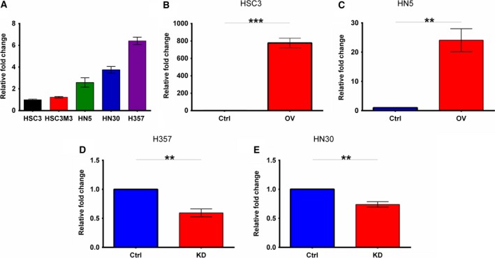
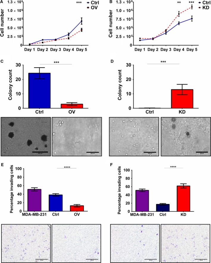
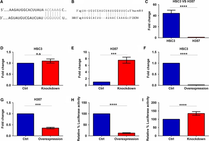
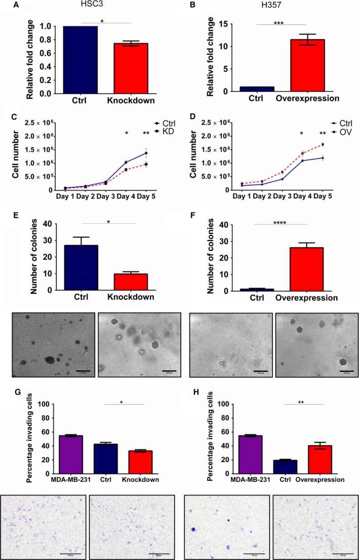
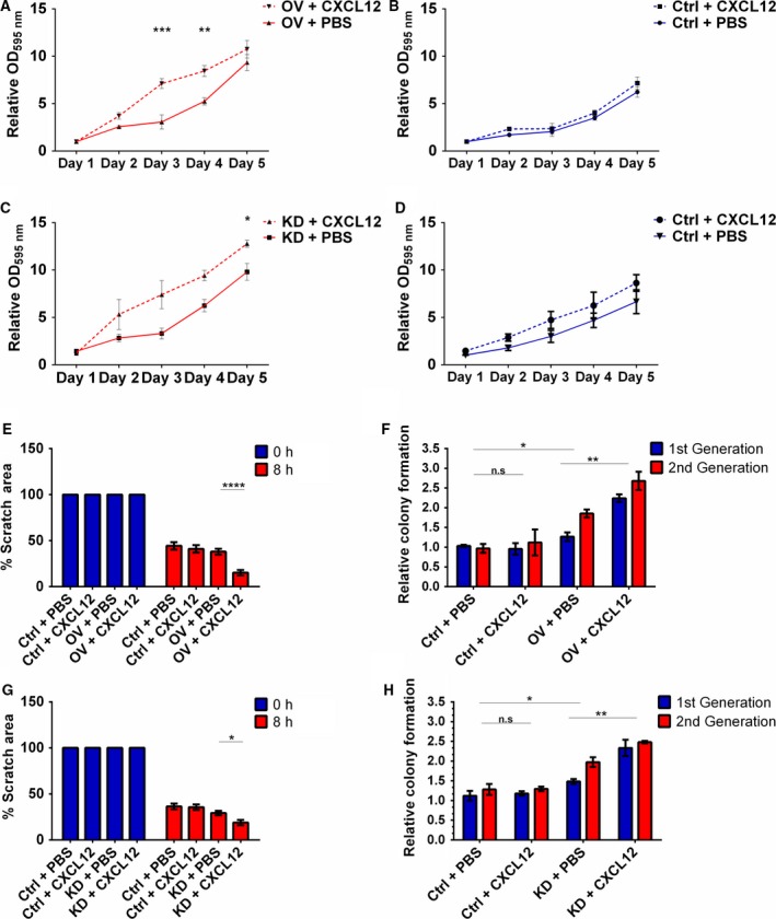
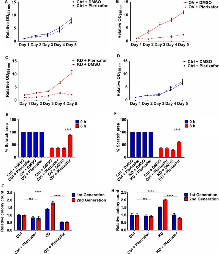
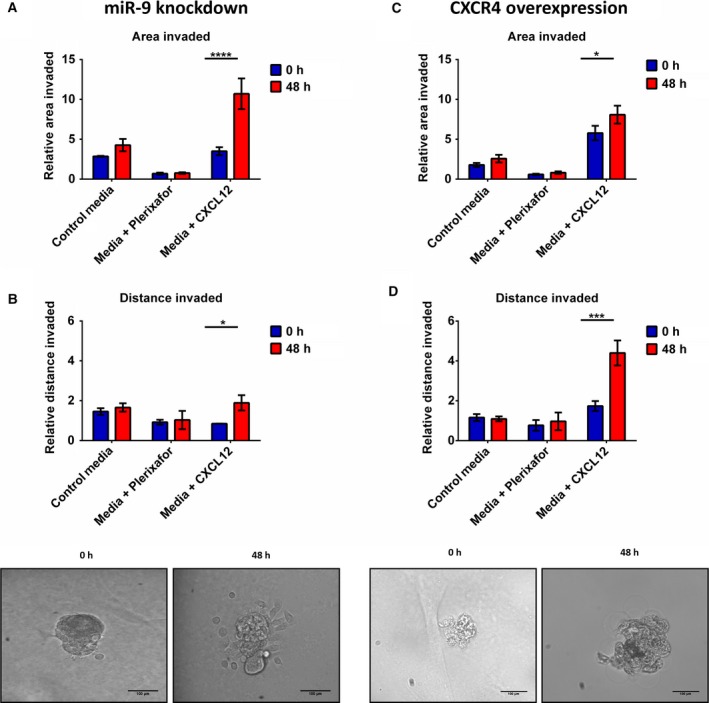
Head and neck squamous cell carcinomas (HNSCC) are associated with poor morbidity and mortality. Current treatment strategies are highly toxic and do not benefit over 50% of patients. There is therefore a crucial need for predictive and/or prognostic biomarkers to allow treatment stratification for individual patients. One class of biomarkers that has recently gained importance are microRNA (miRNA). MiRNA are small, noncoding molecules which regulate gene expression post-transcriptionally. We performed miRNA expression profiling of a cohort of head and neck tumours with known clinical outcomes. The results showed miR-9 to be significantly downregulated in patients with poor treatment outcome, indicating its role as a potential biomarker in HNSCC. Overexpression of miR-9 in HNSCC cell lines significantly decreased cellular proliferation and inhibited colony formation in soft agar. Conversely, miR-9 knockdown significantly increased both these features. Importantly, endogenous CXCR4 expression levels, a known target of miR-9, inversely correlated with miR-9 expression in a panel of HNSCC cell lines tested. Induced overexpression of CXCR4 in low expressing cells increased proliferation, colony formation and cell cycle progression. Moreover, CXCR4-specific ligand, CXCL12, enhanced cellular proliferation, migration, colony formation and invasion in CXCR4-overexpressing and similarly in miR-9 knockdown cells. CXCR4-specific inhibitor plerixafor abrogated the oncogenic phenotype of CXCR4 overexpression as well as miR-9 knockdown. Our data demonstrate a clear role for miR-9 as a tumour suppressor microRNA in HNSCC, and its role seems to be mediated through CXCR4 suppression. MiR-9 knockdown, similar to CXCR4 overexpression, significantly promoted aggressive HNSCC tumour cell characteristics. Our results suggest CXCR4-specific inhibitor plerixafor as a potential therapeutic agent, and miR-9 as a possible predictive biomarker of treatment response in HNSCC.

Keywords: CXCL12; CXCR4; MiR-9; head and neck cancer; plerixafor; tumour invasion.


Figures

Figure 1
Figure 1
miR‐9 expression in a panel of HNSCC cell lines and the modulated cell lines. (A) Relative expression of miR‐9 in six different HNSCC cell lines using qRTPCR normalised against HSC3. (B) HSC3 and (C) HN5 were stably transduced with miR‐9 overexpression vectors, and levels were measured by qRTPCR (D) H357 and (E) HN30 were stably transduced with miR‐9 knockdown vectors, and the efficiency of knockdown was measured by qRTPCR. Data represent mean ± SEM for three independent (= 3) experiments. Asterisks (*) show statistical significance as follows: *< 0.05, **< 0.01, ***< 0.001.
Figure 2
Figure 2
Effect of miR‐9 modulation on proliferation, cell cycle, colony formation and invasion. HSC3 and H357 cell lines were stably transfected using miR‐9 knockdown and overexpression vectors, respectively, and expression was measured by qRTPCR. Cell proliferation of (A) miR‐9 overexpression in HSC3 and (B) miR‐9 knockdown in H357 cells was assessed by generating growth curves over 5 days. The ability of (C) HSC3 miR‐9 overexpression and (D) H357 miR‐9 knockdown cells to form colonies was tested using the soft agar assay. Invasive capacity of (E) HSC3 miR‐9 overexpression and (F) H357 miR‐9 knockdown was assessed using the Transwell Matrigel invasion assay. Representative images were taken at 10× magnification. Data represent mean number of invaded cells through the Matrigel membrane relative to migration through the control membrane. Images are representative of cells fixed and stained on the invasion membrane at 4× magnification. Scale bars = 100 μm Data represent mean ± SEM for three independent (= 3) experiments. Asterisks (*) show statistical significance as follows: *< 0.05, **< 0.01, ***< 0.001, ****< 0.0001.
Figure 3
Figure 3
miR‐9 directly regulates CXCR4 expression. Online computer algorithm (A) TargetScan and (B) miRanda predicting CXCR4 as a miR‐9 target. qRTPCR was performed to compare CXCR4 expression in (C) HSC3 vs H357, (D) HSC3 miR‐9 knockdown, (E) H357 miR‐9 knockdown versus the scrambled controls, whereas (F) HSC3 miR‐9 overexpression and (G) H357 miR‐9 overexpression were compared to the vector control. Luciferase assays confirming miR‐9 interaction with the 3′UTR of CXCR4 were performed for (H) miR‐9 knockdown and (I) miR‐9 overexpression. Data represent mean ±SEM for three independent (= 3) experiments. Asterisks (*) show statistical significance as follows: *< 0.05, **< 0.01, ***< 0.001, ****< 0.0001.
Figure 4
Figure 4
CXCR4 modulations affect cellular proliferation, cell cycle, colony formation and invasion. HSC3 and H357 were stably transfected using (A) CXCR4 knockdown and (B) overexpression vectors and analysed using qRTPCR. Cell proliferation of (C) CXCR4 knockdown and (D) overexpression H357 cells was assessed by generating growth curves over 5 days. The ability of the (E) CXCR4 knockdown and (F) overexpression cells to form colonies was tested using the soft agar assay. Representative images were taken at 10× magnification. Invasive capacity of (G) CXCR4 knockdown and (H) overexpression cells was assessed using the Transwell Matrigel invasion assay. Representative images were taken at 10× magnification. Data represent mean number of invaded cells through the Matrigel membrane relative to migration through the control membrane. Images are representative of cells fixed and stained on the invasion membrane at 4× magnification. Scale bars = 100 μm Data represent mean ± SEM for three independent (= 3) experiments. Asterisks (*) show statistical significance as follows: *< 0.05, **< 0.01, ***< 0.001, ****< 0.0001.
Figure 5
Figure 5
The CXCR4 ligand CXCL12 augments the oncogenic effects of miR‐9 knockdown. Cell proliferation in the presence of CXCL12 was assessed by MTT assay over 5 days in (A) CXCR4‐overexpressing and (B) vector control H357 cells. The effect of CXCL12 on cell proliferation was also assessed by MTT assay in (C) miR‐9 knockdown and (D) scrambled control H357 cells over 5 days. (E) The effect that CXCL12 stimulation of CXCR4 had on migration was examined by scratch assay using CXCR4‐overexpressing H357 cells. (F) Using sphere formation assay, the effect of CXCL12 on colony formation was investigated using CXCR4‐overexpressing H357 cells. (G) Migration of miR‐9 knockdown cells stimulated by CXCL12 was studied using scratch assay. (H) Sphere‐forming capacity of miR‐9 knockdown cells was similarly measured in the presence of CXCL12. Data represent mean ±SEM for three independent (= 3) experiments. Asterisks (*) show statistical significance as follows: *< 0.05, **< 0.01, ***< 0.001, ****< 0.0001.
Figure 6
Figure 6
Plerixafor treatment mimics the tumour‐suppressive effect of miR‐9 via CXCR4 inhibition. Cell proliferation of (A) vector control and (B) CXCR4 overexpressing H357 cells was assessed by MTT assay over 5 days. Proliferation of (C) miR‐9 knockdown and (D) scrambled control H357 cells was also assessed by MTT assay over 5 days. (E) The effect of plerixafor on migration was examined by scratch assay using CXCR4‐overexpressing H357 cells. (F) Using sphere formation assay, the effect on colony formation was investigated using CXCR4‐overexpressing H357 cells. (G) Migration of miR‐9 knockdown cells was studied using scratch assay. (H) Sphere‐forming capacity of miR‐9 knockdown cells was similarly measured in the presence of plerixafor. Data represent mean ±SEM for three independent (= 3) experiments. Asterisks (*) show statistical significance as follows: *< 0.05, **< 0.01, ***< 0.001, ****< 0.0001.
Figure 7
Figure 7
miR‐9 and CXCR4 regulate invasion under anchorage‐independent conditions. Invasion of miR‐9 knockdown cells was quantitated using imagej software. Invasion of miR‐9 knockdown cells was calculated by measuring the (A) total area invaded by the spheroid and (B) the longest invasive distance from the spheroid. Invasion of CXCR4‐overexpressing cell was also investigated by (C) total area invaded and (D) longest invasive distance. Data represent mean ±SEM for three independent (= 3) experiments. Scale bars = 100 μm Asterisks (*) show statistical significance as follows: *< 0.05, **< 0.01, ***< 0.001, ****< 0.0001.

References

    1. Albert S, Riveiro ME, Halimi C, Hourseau M, Couvelard A, Serova M, Barry B, Raymond E and Faivre S (2013) Focus on the role of the CXCL12/CXCR4 chemokine axis in head and neck squamous cell carcinoma. Head Neck 35, 1819–1828. - PubMed
    1. Anonymous (2007) Plerixafor: AMD 3100, AMD3100, JM 3100, SDZ SID 791. Drugs R D 8, 113–119. - PubMed
    1. Balkwill F (2004) Cancer and the chemokine network. Nat Rev Cancer 4, 540–550. - PubMed
    1. Bandres E, Agirre X, Bitarte N, Ramirez N, Zarate R, Roman‐Gomez J, Prosper F and Garcia‐Foncillas J (2009) Epigenetic regulation of microRNA expression in colorectal cancer. Int J Cancer 125, 2737–2743. - PubMed
    1. Bazzoni F, Rossato M, Fabbri M, Gaudiosi D, Mirolo M, Mori L, Tamassia N, Mantovani A, Cassatella MA and Locati M (2009) Induction and regulatory function of miR‐9 in human monocytes and neutrophils exposed to proinflammatory signals. Proc Natl Acad Sci U S A 106, 5282–5287. - PMC - PubMed

Publication types

MeSH terms

LinkOut - more resources