Thrombin modifies growth, proliferation and apoptosis of human colon organoids: a protease-activated receptor 1- and protease-activated receptor 4-dependent mechanism
- PMID: 29959891
- PMCID: PMC6109216
- DOI: 10.1111/bph.14430
Thrombin modifies growth, proliferation and apoptosis of human colon organoids: a protease-activated receptor 1- and protease-activated receptor 4-dependent mechanism
Abstract
Background and purpose: Thrombin is massively released upon tissue damage associated with bleeding or chronic inflammation. The effects of this thrombin on tissue regrowth and repair has been scarcely addressed and only in cancer cell lines. Hence, the purpose of the present study was to determine thrombin's pharmacological effects on human intestinal epithelium growth, proliferation and apoptosis, using three-dimensional cultures of human colon organoids.
Experimental approach: Crypts were isolated from human colonic resections and cultured for 6 days, forming human colon organoids. Cultured organoids were exposed to 10 and 50 mU·mL-1 of thrombin, in the presence or not of protease-activated receptor (PAR) antagonists. Organoid morphology, metabolism, proliferation and apoptosis were followed.
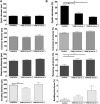
Key results: Thrombin favoured organoid maturation leading to a decreased number of immature cystic structures and a concomitant increased number of larger structures releasing cell debris and apoptotic cells. The size of budding structures, metabolic activity and proliferation were significantly reduced in organoid cultures exposed to thrombin, while apoptosis was dramatically increased. Both PAR1 and PAR4 antagonists inhibited apoptosis regardless of thrombin doses. Thrombin-induced inhibition of proliferation and metabolic activity were reversed by PAR4 antagonist for thrombin's lowest dose and by PAR1 antagonist for thrombin's highest dose.
Conclusions and implications: Overall, our data suggest that the presence of thrombin in the vicinity of human colon epithelial cells favours their maturation at the expense of their regenerative capacities. Our data point to thrombin and its two receptors PAR1 and PAR4 as potential molecular targets for epithelial repair therapies.
© 2018 The Authors. British Journal of Pharmacology published by John Wiley & Sons Ltd on behalf of British Pharmacological Society.
Figures


Similar articles
-
Protease-activated receptor 4 uses anionic residues to interact with alpha-thrombin in the absence or presence of protease-activated receptor 1.Biochemistry. 2008 Dec 16;47(50):13279-86. doi: 10.1021/bi801334s. Biochemistry. 2008. PMID: 19053259
-
Neuroprotective signal transduction in model motor neurons exposed to thrombin: G-protein modulation effects on neurite outgrowth, Ca(2+) mobilization, and apoptosis.J Neurobiol. 2001 Aug;48(2):87-100. J Neurobiol. 2001. PMID: 11438939
-
Protease-activated receptors 1 and 4 mediate activation of human platelets by thrombin.J Clin Invest. 1999 Mar;103(6):879-87. doi: 10.1172/JCI6042. J Clin Invest. 1999. PMID: 10079109 Free PMC article.
-
Therapeutic potential of protease-activated receptor-1 antagonists.Expert Opin Investig Drugs. 2003 Feb;12(2):209-21. doi: 10.1517/13543784.12.2.209. Expert Opin Investig Drugs. 2003. PMID: 12556215 Review.
-
Thrombin stories in the gut.Biochimie. 2024 Nov;226:107-112. doi: 10.1016/j.biochi.2024.03.007. Epub 2024 Mar 22. Biochimie. 2024. PMID: 38521125 Review.
Cited by
-
Unveiling how mitotic spindle orientation in 3D human colon organoids affects matrix displacements through a 4D study using DVC.Sci Rep. 2025 Jul 1;15(1):22052. doi: 10.1038/s41598-025-04156-4. Sci Rep. 2025. PMID: 40595987 Free PMC article.
-
Epithelial production of elastase is increased in inflammatory bowel disease and causes mucosal inflammation.Mucosal Immunol. 2021 May;14(3):667-678. doi: 10.1038/s41385-021-00375-w. Epub 2021 Mar 5. Mucosal Immunol. 2021. PMID: 33674762 Free PMC article.
-
Active thrombin produced by the intestinal epithelium controls mucosal biofilms.Nat Commun. 2019 Jul 19;10(1):3224. doi: 10.1038/s41467-019-11140-w. Nat Commun. 2019. PMID: 31324782 Free PMC article.
-
Sexual dimorphism in PAR2-dependent regulation of primitive colonic cells.Biol Sex Differ. 2019 Sep 6;10(1):47. doi: 10.1186/s13293-019-0262-6. Biol Sex Differ. 2019. PMID: 31492202 Free PMC article.
-
Protease-Activated Receptors - Key Regulators of Inflammatory Bowel Diseases Progression.J Inflamm Res. 2021 Dec 29;14:7487-7497. doi: 10.2147/JIR.S335502. eCollection 2021. J Inflamm Res. 2021. PMID: 35002281 Free PMC article. Review.
References
-
- Cenac N, Altier C, Motta JP, d'Aldebert E, Galeano S, Zamponi GW et al (2010). Potentiation of TRPV4 signalling by histamine and serotonin: an important mechanism for visceral hypersensitivity. Gut 59: 481–488. - PubMed
-
- Cenac N, Cellars L, Steinhoff M, Andrade‐Gordon P, Hollenberg MD, Wallace JL et al (2005). Proteinase‐activated receptor‐1 is an anti‐inflammatory signal for colitis mediated by a type 2 immune response. Inflamm Bowel Dis 11: 792–798. - PubMed
Publication types
MeSH terms
Substances
LinkOut - more resources
Full Text Sources
Other Literature Sources
Research Materials

