

Identification of 7α,24-dihydroxy-3-oxocholest-4-en-26-oic and 7α,25-dihydroxy-3-oxocholest-4-en-26-oic acids in human cerebrospinal fluid and plasma
- PMID: 29960034
- PMCID: PMC6171785
- DOI: 10.1016/j.biochi.2018.06.020
Identification of 7α,24-dihydroxy-3-oxocholest-4-en-26-oic and 7α,25-dihydroxy-3-oxocholest-4-en-26-oic acids in human cerebrospinal fluid and plasma
Abstract
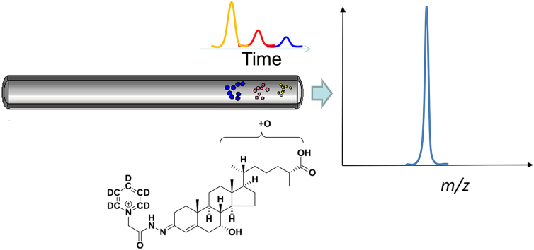
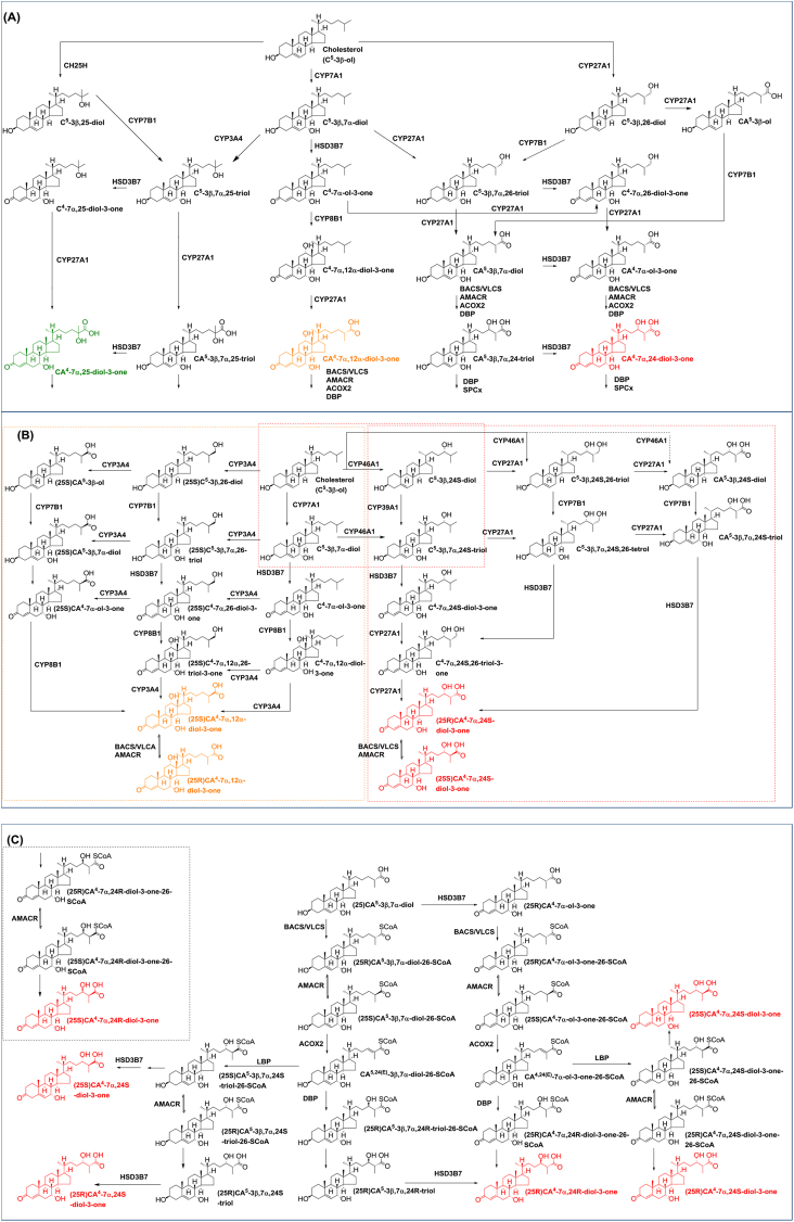
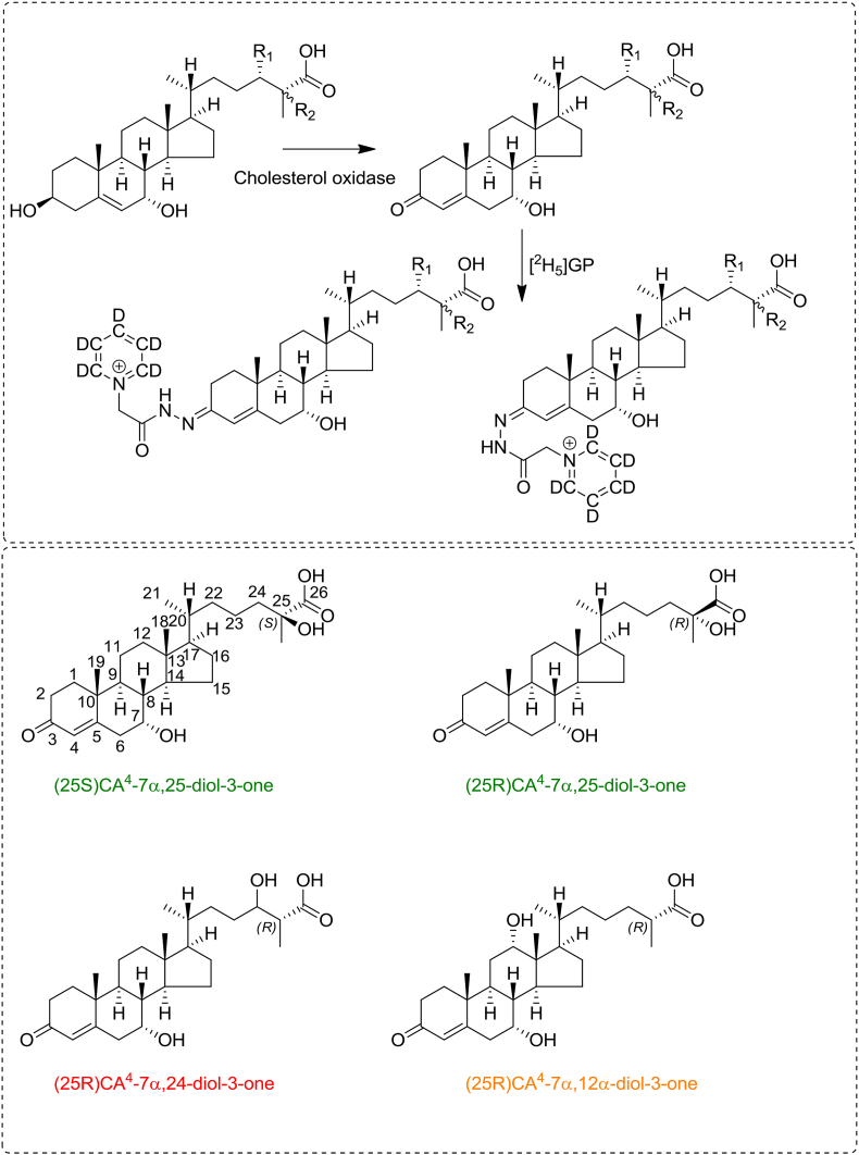
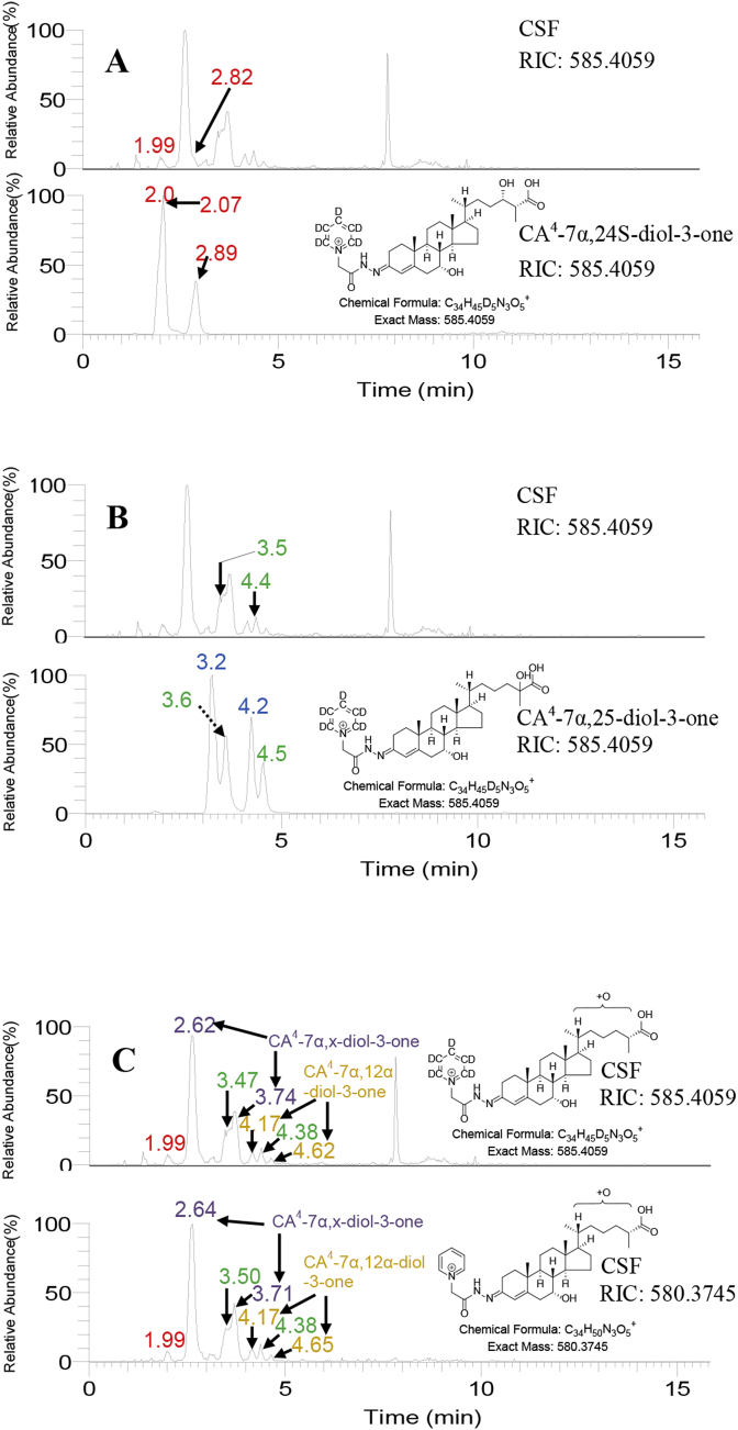
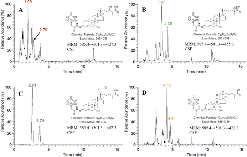
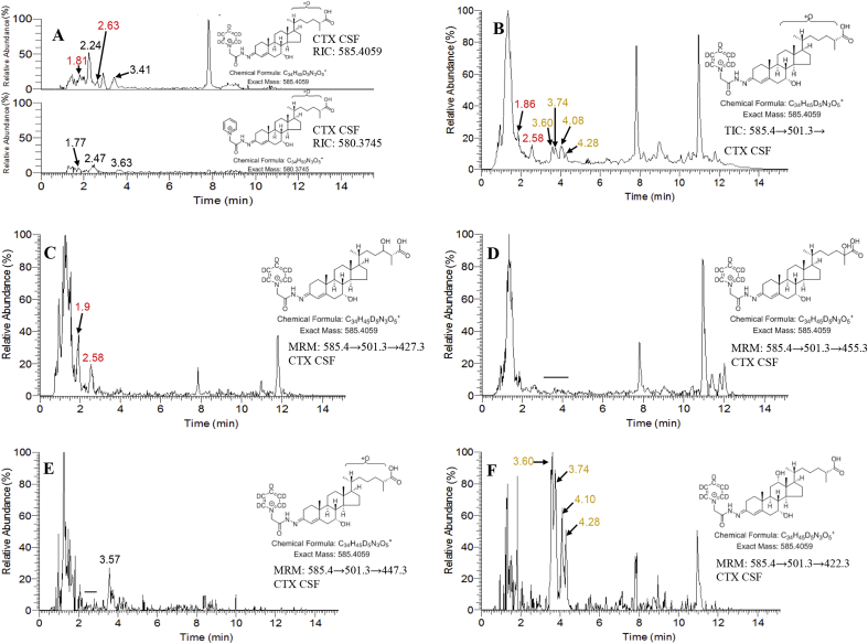
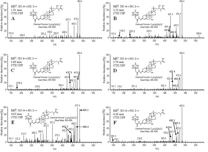
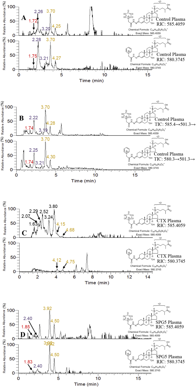
Dihydroxyoxocholestenoic acids are intermediates in bile acid biosynthesis. Here, using liquid chromatography - mass spectrometry, we confirm the identification of 7α,24-dihydroxy-3-oxocholest-4-en-26-oic and 7α,25-dihydroxy-3-oxocholest-4-en-26-oic acids in cerebrospinal fluid (CSF) based on comparisons to authentic standards and of 7α,12α-dihydroxy-3-oxocholest-4-en-26-oic and 7α,x-dihydroxy-3-oxocholest-4-en-26-oic (where hydroxylation is likely on C-22 or C-23) based on exact mass measurement and multistage fragmentation. Surprisingly, patients suffering from the inborn error of metabolism cerebrotendinous xanthomatosis, where the enzyme CYP27A1, which normally introduces the (25 R)26-carboxylic acid group to the sterol side-chain, is defective still synthesise 7α,24-dihydroxy-3-oxocholest-4-en-26-oic acid and also both 25 R- and 25 S-epimers of 7α,12α-dihydroxy-3-oxocholest-4-en-26-oic acid. We speculate that the enzymes CYP46A1 and CYP3A4 may have C-26 carboxylase activity to generate these acids. In patients suffering from hereditary spastic paraplegia type 5 the CSF concentrations of the 7α,24- and 7α,25-dihydroxy acids are reduced, suggesting an involvement of CYP7B1 in their biosynthesis in brain.
Keywords: Bile acid; Brain; Cerebrotendinous xanthomatosis; Cytochrome P450; Hereditary spastic paraplegia type 5; Oxysterol.
Copyright © 2018 The Authors. Published by Elsevier B.V. All rights reserved.
Figures

Similar articles
-
Additional pathways of sterol metabolism: Evidence from analysis of Cyp27a1-/- mouse brain and plasma.Biochim Biophys Acta Mol Cell Biol Lipids. 2019 Feb;1864(2):191-211. doi: 10.1016/j.bbalip.2018.11.006. Epub 2018 Nov 22. Biochim Biophys Acta Mol Cell Biol Lipids. 2019. PMID: 30471425 Free PMC article.
-
Cholestenoic acids regulate motor neuron survival via liver X receptors.J Clin Invest. 2014 Nov;124(11):4829-42. doi: 10.1172/JCI68506. Epub 2014 Oct 1. J Clin Invest. 2014. PMID: 25271621 Free PMC article.
-
Cerebrospinal fluid steroidomics: are bioactive bile acids present in brain?J Biol Chem. 2010 Feb 12;285(7):4666-79. doi: 10.1074/jbc.M109.086678. Epub 2009 Dec 7. J Biol Chem. 2010. PMID: 19996111 Free PMC article.
-
Cerebrotendinous xanthomatosis: an inborn error in bile acid synthesis with defined mutations but still a challenge.Biochem Biophys Res Commun. 2010 May 21;396(1):46-9. doi: 10.1016/j.bbrc.2010.02.140. Biochem Biophys Res Commun. 2010. PMID: 20494109 Review.
-
Cytochrome P450s in the synthesis of cholesterol and bile acids--from mouse models to human diseases.FEBS J. 2012 May;279(9):1516-33. doi: 10.1111/j.1742-4658.2011.08432.x. Epub 2011 Dec 22. FEBS J. 2012. PMID: 22111624 Review.
Cited by
-
Cholesterol accessibility at the ciliary membrane controls hedgehog signaling.Elife. 2019 Oct 30;8:e50051. doi: 10.7554/eLife.50051. Elife. 2019. PMID: 31657721 Free PMC article.
-
Cholesterol metabolism: from lipidomics to immunology.J Lipid Res. 2022 Feb;63(2):100165. doi: 10.1016/j.jlr.2021.100165. Epub 2021 Dec 22. J Lipid Res. 2022. PMID: 34953867 Free PMC article. Review.
-
The Biosynthesis of Enzymatically Oxidized Lipids.Front Endocrinol (Lausanne). 2020 Nov 19;11:591819. doi: 10.3389/fendo.2020.591819. eCollection 2020. Front Endocrinol (Lausanne). 2020. PMID: 33329396 Free PMC article. Review.
-
Lipid metabolic pathways converge in motor neuron degenerative diseases.Brain. 2020 Apr 1;143(4):1073-1087. doi: 10.1093/brain/awz382. Brain. 2020. PMID: 31848577 Free PMC article. Review.
-
Oxysterols as lipid mediators: Their biosynthetic genes, enzymes and metabolites.Prostaglandins Other Lipid Mediat. 2020 Apr;147:106381. doi: 10.1016/j.prostaglandins.2019.106381. Epub 2019 Nov 4. Prostaglandins Other Lipid Mediat. 2020. PMID: 31698146 Free PMC article. Review.
References
-
- Russell D.W. The enzymes, regulation, and genetics of bile acid synthesis. Annu. Rev. Biochem. 2003;72:137–174. - PubMed
-
- Vaz F.M., Ferdinandusse S. Bile acid analysis in human disorders of bile acid biosynthesis. Mol. Aspect. Med. 2017;56:10–24. - PubMed
-
- Schols L., Rattay T.W., Martus P., Meisner C., Baets J., Fischer I., Jagle C., Fraidakis M.J., Martinuzzi A., Saute J.A., Scarlato M., Antenora A., Stendel C., Hoflinger P., Lourenco C.M., Abreu L., Smets K., Paucar M., Deconinck T., Bis D.M., Wiethoff S., Bauer P., Arnoldi A., Marques W., Jardim L.B., Hauser S., Criscuolo C., Filla A., Zuchner S., Bassi M.T., Klopstock T., De Jonghe P., Bjorkhem I., Schule R. Hereditary spastic paraplegia type 5: natural history, biomarkers and a randomized controlled trial. Brain. 2017;140:3112–3127. - PMC - PubMed
MeSH terms
Substances
Grants and funding
LinkOut - more resources
Full Text Sources
Other Literature Sources
Research Materials
