. 2018 Oct 30:675:15-26.
doi: 10.1016/j.gene.2018.06.092. Epub 2018 Jun 28.

Alternative splicing of (ppp1r12a/mypt1) in zebrafish produces a novel myosin phosphatase targeting subunit

Affiliations

Alternative splicing of (ppp1r12a/mypt1) in zebrafish produces a novel myosin phosphatase targeting subunit

Andrew LaFlamme et al. Gene. .

Abstract

Myosin phosphatase is an evolutionarily conserved regulator of actomyosin contractility, comprised of a regulatory subunit (Mypt1), and a catalytic subunit (PP1). Zebrafish has become an ideal model organism for the study of the genetic and cell physiological role of the myosin phosphatase in morphogenesis and embryonic development. We identified and characterized a novel splice variant of Mypt1 (ppp1r12a-tv202) from zebrafish, which is widely expressed during early embryonic development. Importantly, mutant alleles and antisense morpholinos that have been used to demonstrate the important role of Mypt1 in early development, not only disrupt the longer splice variants, but also tv202. The protein product of ppp1r12a-tv202 (Mypt1-202) contains the PP1-binding N-terminus, but lacks the regulatory C-terminus, which contains two highly conserved inhibitory phosphorylation sites. We observed that the protein product of tv202 assembled a constitutively active myosin phosphatase uninhibited by kinases such as Zipk. Thus, we propose that Mypt1-202 plays an important role in maintaining baseline Mlc2 dephosphorylation and actomyosin relaxation during early zebrafish development.

Keywords: Actomyosin; Convergent extension; Gastrulation; Myosin phosphatase; Mypt1; PP1; Splicing; Zebrafish.


Conflict of interest statement

Conflict of interest

The authors declare no conflict of interest

Figures

Figure 1.
Figure 1.
(A) A schematic diagram MYPT1 splice variants tv201 (top) and tv202 (bottom). 100 kb of genomic sequence from 4:21765599 to 4:21865598 is represented. The first 11 exons of tv201 and tv202 are similar. The 3’ splice site in tv201 is bypassed in tv202, yielding a longer exon 12 and an alternative final exon 13. Protein coding regions are in black, non-coding regions are gray. Tv201 contains 26 exons. (B) A diagram of the domain architecture of the protein-product for ppplrl2a-F201 and the protein-product of ppp1r12a-tv202. The conserved PP1 binding domain consisting of the KVRF-motif (aa 35–38) and subsequent and ankyrin repeats are labelled. The inhibitory phosphorylation sites T725 and T877 and leucine zipper domain are shown on tv201. The unique sequence found in the protein product of TV202 is shown in the inset.
Figure 2.
Figure 2.
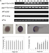
Expression of ppp1r12a splice variants during early embryonic development in zebrafish. (A) Transcript specific primers were used to detect ppp1r12a-tv202 or to detect the long transcript variants tv-201 and tv-202, labelled tv-long. Embryos were collected immediately after fertilization (0 hpf), 64-cell stage (2 hpf), sphere stage (4 hpf), shield stage (6 hpf), bud stage (10 hpf), 6 somite stage (12 hpf), 10 somite stage (14 hpf), 22 somite stage (20 hpf), Prim-5 stage (24 hpf), long-pec stage (48 hpf) and protruding-month stage (72 hpf). Amplification of eF1a and total RNA without addition of reverse transcriptase were used as controls. The PCR products amplified by tv-long, tv202 and eF1a primers were 769 bp, 206 bp and 720 bp in length respectively. (B-D) The spatial expression of ppp1r12a-tv202 was analyzed by whole-mount in situ hybridization using a probe targeted at the unique regions of exon 12 and 13 in tv202 at the 64 cell stage (B), bud stage (C) and 24 hpf (D). Quantitative analysis of mRNA expression levels of long-tv (E) and tv202 (F) during zebrafish development. Values are means of three biological replicates performed in duplicate and error bars are standard deviation. All values are normalized the eF1a and reported as % of expression of the long-tv expression at 0 hpf.
Figure 3.
Figure 3.
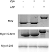
Mypt1 202 is not subject to inhibitory phosphorylation. Zebrafish Zipk immunoprecipitated from HEK 293T cells was used to phosphorylate purified GST- Mypt1, GST-Mlc2 or Myptl-202 immunoprecipitated from HEK 293T cells. Phosphorylation was detected by band shift using a phos-tag SDS-PAGE. These experiments were repeated a total of five times.
Figure 4.
Figure 4.
Subcellular localization of Myptl-202. HeLa cells were transfected with either GFP-Mypt 1–202 (A, B), GFP-Mypt1-202 and flag- PPlß. (C, D), GFP-Myptl-Δ (E, F), GFP- Myptl- Δ and flag-PPlß (G,H), GFP-alone (I, J). Untransfected HeLa cells were treated with the myosin inhibitor blebbistatin (L) or a DMSO control (K). All cells were fixed and stained with DAPI and Alexa 568-phalloidin and imaged with confocal microscopy. Black and white images show GFP localization, while color images are a merge of DAPI (blue), GFP (green) and phalloidin (red). White bar indicates 20 pm. Representative images are displayed from a minimum of three biological replicates and 30 individual cells per condition.
Figure 5.
Figure 5.
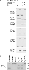
Mypt1-202 binds PPlß forming an active myosin phosphatase complex. (A) Myc-tagged Mypt1-202 or Mypt1-Δ and GFP-tagged PPlß or GFP were expressed in HEK293T cells and immunoprecipitated with either anti-myc or anti-GFP antibodies. The IPs were immunoblotted with anti-GFP antibodies and anti-myc antibodies. (B) GST-Mlc2 was phosphorylated in vitro using Zipk (phospho) or left unphosphorylated (unphospho). The prephosphorylated Mlc2 was dephosphorylated by Myptl-PPl complexes immunoprecipitated from HEK293T cell lysates or using controls of treatment with beads from a myc-IP from untransfected cells (mock IP). The Myptl complexes were collected from cells transfected with Myptl-202 and PPlß, Myptl-Δ and PPlß or a non-PPl binding mutant Myptl (1–300 RARA) and PPlß. Dephosphorylation (0), mono (1) and di-phosphorylation (2) was detected by band shift using a phos-tag SDS-PAGE gel. All biochemical analyses were performed a minimum of three times.
Figure 6.
Figure 6.
Myptl-202 and PPlß assemble a myosin phosphatase complex and regulate the actin cytoskeleton. HeLa cells were transfected with either myc-Mypt1-202 and GFP (A, B), myc-Mypt1A (C, D), myc-Myptl-202 and GFP-PPlß (E, F), myc-Mypt1A and GFP- PPlß (G, H), GFP alone (I, J), or GFP-PPlß (K, L). All cells were fixed and stained with DAPI and Alexa 568-phalloidin to stain the nucleus and actin cytoskeleton, respectively, and imaged with confocal microscopy. Black and white images show phalloidin staining, while color images are a merge of DAPI (blue), GFP (green) and phalloidin (red). White bar indicates 20 pm. Representative cells are shown from a minimum of three biological replicates.
Figure 7.
Figure 7.
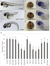
Myptl-202 expression can resuce Myptl loss-of-function in zebrafish embryos. Lateral views of representative 48 hpf zebrafish embryos injected with (A) control morpholino, (B) 1.0 ng ppp1r12a MO, (C) a partially rescued embryo injected with 25 pg ppp1r12a-tv202 mRNA and 1.0 ng of ppp1r12a MO. (D-F) Embryos at bud stage stained with hggl (to mark the prechordal plate), shh (midline), pax2.1 (midbrain- hindbrain boundary) and dix3 (neural plate) after injection of (D) control morpholino, (E) ppplrl2aMO or(F)ppp1rl2a-tv202 mRNA and ppp1r12a MO. (G) Quantification of the truncated body axis phenotype in morphant and mRNA injected embryos. Error bars are standard error and a* indicates a statistically significant (p<0.05) difference from control and a # indicates a statistically significant (p<0.05) rescue compared to the corresponding morpholino injected embryos. Injections were performed a minimum of three times with 50 total embryos measured. Embryos were collected by spawning hétérozygotie hi2653 adult zebrafish (I and J) or from WT controls (H). Mutant embryos were injected with 25 pg of ppp1r12a-tv202 mRNA at the one cell stage (I). Embryos were collected at 36 hpf and probed with proxl to mark the liver primordium (Marked with an arrowhead). Liver staining for proxl was detected in 68 of 68 wild-type embryos, 57 of 77 uninjected embryos from the hétérozygotie cross and 89/91 rescued embryos.
Figure 8.
Figure 8.
Overexpression of Myptl-202 in zebrafish embryos results in constitutive dephosphorylation of Mlc2. Lateral views of representative 48 hpf zebrafish embryos injected with (A) 200 pg myc-long-mypt1 mRNA or (C) 100 pg myc-ppp1r12a-tv202 mRNA. Embryos at bud stage stained with hggl (to mark the prechordal plate), shh(midline),pax2.1 (midbrain-hindbrain boundary) and dix 3 (neural plate) after injection of (B) 200 pg myc-long-mypt1 mRNA or (D) 100 pg myc-ppp1r12a-tv202 mRNA. A representative field of presomitic and notochordal mesoderm at the bud stage in control embryos (E), embryos injected with 200 pg myc-long-myptl mRNA (F) or 100 pg ppp1r12ci-tv202 mRNA (G). (H) The actomyosin driven elongation of mesodermal cells was determined by calculating the length width ratio (y-axis). The crossed bars indicate cells with the long axis length and the short axis width. Error bars are standard error and a * indicates a statistically significant difference from control. All calculations were made on 75 cells from 3–5 separate embryos. Statistical significance was calculated using a one-factor ANOVA with Tukey post hoc analysis and is defined as p < 0.05. (I) Embryo lysates from 25 control embryos or 25 embryos injected with either myc-long-mypt1 mRNA or myc-ppp1r12a-tv202 mRNA were analyzed by western blot using anti-myc, anti-phospho-Mlc2 or anti-Mlc2 antibodies. This biochemical analysis was performed four times.

Similar articles

Cited by

References

    1. Landsverk ML & Epstein HF Genetic analysis of myosin II assembly and organization in model organisms. Cellular and Molecular Life Sciences 62, 2270–2282 (2005). - PMC - PubMed
    1. Vicente-Manzanares M, Ma X, Adelstein RS & Horwitz AR Non-muscle myosin II takes centre stage in cell adhesion and migration. Nature Reviews Molecular Cell Biology 10, 778–790 (2009). - PMC - PubMed
    1. Hirano K, Derkach DN, Hirano M, Nishimura J & Kanaide H Protein kinase network in the regulation of phosphorylation and dephosphorylation of smooth muscle myosin light chain. Molecular and Cellular Biochemistry 248, 105–114(2003). - PubMed
    1. Grassie ME, Moffat LD, Walsh MP & MacDonald JA The myosin phosphatase targeting protein (MYPT) family: A regulated mechanism for achieving substrate specificity of the catalytic subunit of protein phosphatase type 1δ. Archives of Biochemistry and Biophysics 510, 147–159 (2011). - PubMed
    1. Ito M, Nakano T, Erdodi F & Hartshome DJ Myosin phosphatase: Structure, regulation and function. Molecular and Cellular Biochemistry 259, 197–209 (2004). - PubMed

MeSH terms

LinkOut - more resources