

Cousins at work: How combining medical with optical imaging enhances in vivo cell tracking
- PMID: 29960079
- PMCID: PMC6593261
- DOI: 10.1016/j.biocel.2018.06.008
Cousins at work: How combining medical with optical imaging enhances in vivo cell tracking
Abstract
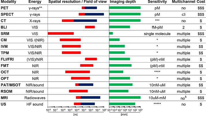
Microscopy and medical imaging are related in their exploitation of electromagnetic waves, but were developed to satisfy differing needs, namely to observe small objects or to look inside subjects/objects, respectively. Together, these techniques can help elucidate complex biological processes and better understand health and disease. A current major challenge is to delineate mechanisms governing cell migration and tissue invasion in organismal development, the immune system and in human diseases such as cancer where the spatiotemporal tracking of small cell numbers in live animal models is extremely challenging. Multi-modal multi-scale in vivo cell tracking integrates medical and optical imaging. Fuelled by basic research in cancer biology and cell-based therapeutics, it has been enabled by technological advances providing enhanced resolution, sensitivity and multiplexing capabilities. Here, we review which imaging modalities have been successfully used for in vivo cell tracking and how this challenging task has benefitted from combining macroscopic with microscopic techniques.
Keywords: Cancer metastasis; Cell therapy; Microscopy; Reporter genes; Whole-body imaging.
Copyright © 2018. Published by Elsevier Ltd.
Figures
References
-
- Alanentalo T., Loren C.E., Larefalk A., Sharpe J., Holmberg D., Ahlgren U. High-resolution three-dimensional imaging of islet-infiltrate interactions based on optical projection tomography assessments of the intact adult mouse pancreas. J. Biomed. Opt. 2008;13:054070. - PubMed
Publication types
MeSH terms
Grants and funding
LinkOut - more resources
Full Text Sources
Other Literature Sources
Medical
