. 2018 Jun 30;7(13):e008757.
doi: 10.1161/JAHA.118.008757.

Dietary Docosahexaenoic Acid Reduces Oscillatory Wall Shear Stress, Atherosclerosis, and Hypertension, Most Likely Mediated via an IL-1-Mediated Mechanism

Affiliations

Dietary Docosahexaenoic Acid Reduces Oscillatory Wall Shear Stress, Atherosclerosis, and Hypertension, Most Likely Mediated via an IL-1-Mediated Mechanism

Mabruka A Alfaidi et al. J Am Heart Assoc. .

Abstract

Background: Hypertension is a complex condition and a common cardiovascular risk factor. Dietary docosahexaenoic acid (DHA) modulates atherosclerosis and hypertension, possibly via an inflammatory mechanism. IL-1 (interleukin 1) has an established role in atherosclerosis and inflammation, although whether IL-1 inhibition modulates blood pressure is unclear.

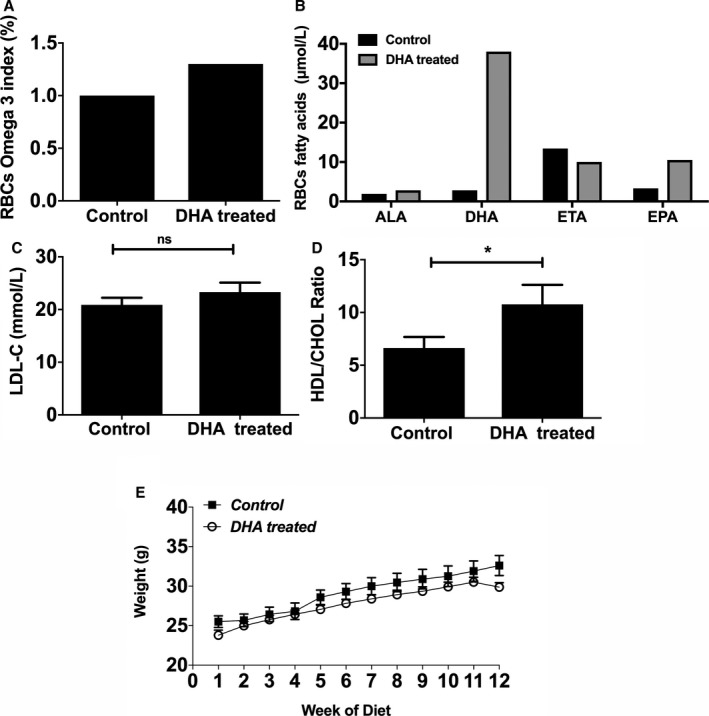
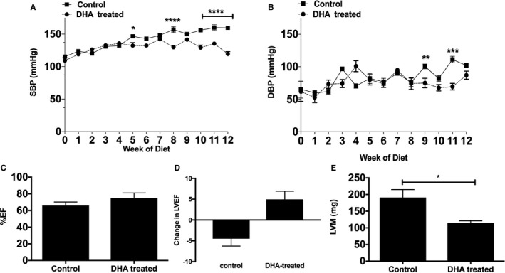
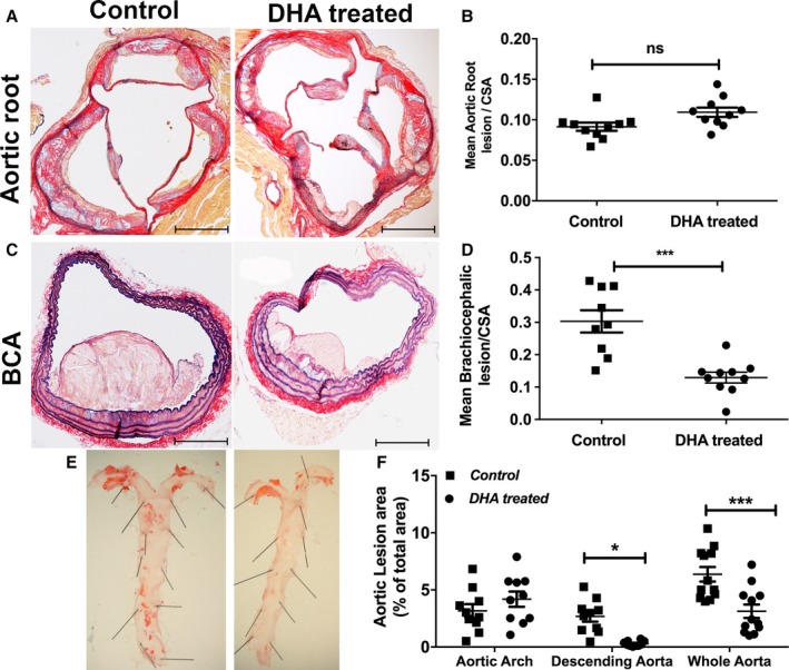
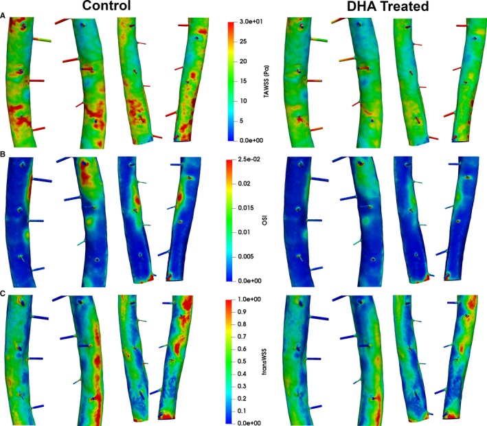
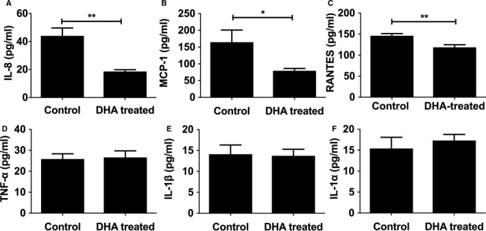
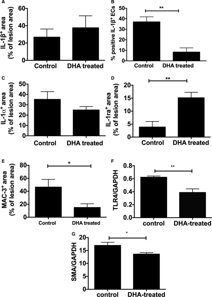
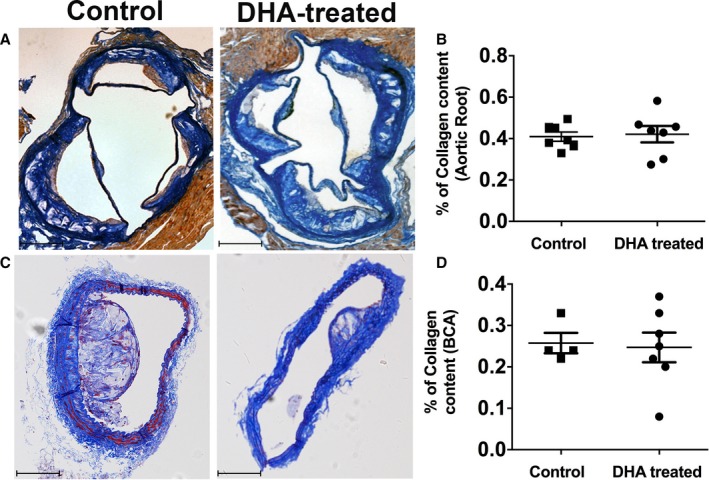
Methods and results: Male apoE-/- (apolipoprotein E-null) mice were fed either a high fat diet or a high fat diet plus DHA (300 mg/kg per day) for 12 weeks. Blood pressure and cardiac function were assessed, and effects of DHA on wall shear stress and atherosclerosis were determined. DHA supplementation improved left ventricular function, reduced wall shear stress and oscillatory shear at ostia in the descending aorta, and significantly lowered blood pressure compared with controls (119.5±7 versus 159.7±3 mm Hg, P<0.001, n=4 per group). Analysis of atheroma following DHA feeding in mice demonstrated a 4-fold reduction in lesion burden in distal aortas and in brachiocephalic arteries (P<0.001, n=12 per group). In addition, DHA treatment selectively decreased plaque endothelial IL-1β (P<0.01).

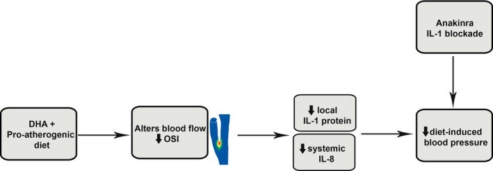
Conclusions: Our findings revealed that raised blood pressure can be reduced by inhibiting IL-1 indirectly by administration of DHA in the diet through a mechanism that involves a reduction in wall shear stress and local expression of the proinflammatory cytokine IL-1β.

Keywords: docosahexaenoic acid; endothelium; hypertension; inflammation; interleukin 1; wall shear stress.


Figures

Figure 1
Figure 1
DHA feeding of apoE−/− (apolipoprotein E–null) mice induces changes in RBC ω‐3 fatty acid compositions without any significant effect on their body weights. A, The ω‐3 index (percentage) is enhanced in DHA‐fed mice compared with controls. The apoE−/− mice were fed an HFD alone (control) or an HFD and DHA (300 mg/kg per day) for 12 weeks. Data are from pooled blood of n=4 mice per group. Blood was sampled at the experimental end point. B, Fatty acid composition in RBCs of DHA‐fed mice compared with controls (μmol/L; pooled blood from 4 mice per group). C, Plasma LDL‐C (in mmol/L) and (D) HDL/CHOL ratio, measured enzymatically (n=10 per group). E, Body weights of the mice (in g) were recorded weekly. Data are shown as mean±SEM, analyzed by Student t test (C and D) or 2‐way ANOVA followed by Tukey test (E), *P<0.05. ALA indicates α‐linolenic acid (18:3n‐3); DHA, docosahexaenoic acid (22:6n‐3); EPA, eicosapentaenoic acid (20:5n‐3); ETA, eicosatetraenoic acid (20:4n‐3); HDL/CHOL, high‐density lipoprotein/total cholesterol; HFD, high‐fat diet; LDL‐C, low‐density lipoprotein cholesterol; RBC, red blood cell.
Figure 2
Figure 2
DHA significantly reduces HFD induced hypertension in apoE−/− (apolipoprotein E–null) mice. A, SBP and (B) DBP were measured in freely moving apoE−/− mice fed either HFD alone (control) or HFD and DHA (DHA treated) for 12 weeks (n=4 per group) using a tail‐cuff system (see Methods for details). All data are expressed as mean±SEM, analyzed by 2‐way ANOVA and Tukey posttest, *P<0.05, **P<0.01, ***P<0.001, ****P<0.0001. Effects of DHA feeding on (C) left ventricular EF (%EF) and change in (D) EF and (E) LVM (in mg). Echocardiographic data were measured in anesthetized mice and are presented as mean±SEM (n=4 per group), analyzed by unpaired Student t test, *P<0.05. DBP, diastolic blood pressure; DHA, docosahexaenoic acid (22:6n‐3); EF, ejection fraction; HFD, high‐fat diet; LVM, left ventricular mass; SBP, systolic blood pressure.
Figure 3
Figure 3
Differential effects of DHA feeding on lesion development in different areas of the vascular beds. Male apoE−/− (apolipoprotein E–null) mice aged 8 weeks were fed a Western‐type HFD alone (control) or an HFD and jelly containing DHA (DHA treated; 300 mg/kg per day) daily over 12 weeks. A, Representative images of aortic roots stained with AB/EVG after 12 weeks of feeding (n=12 per group). Scale bars=100 μm. B, Mean lesion area of aortic root sections, normalized to CSA (n=12). Data are mean±SEM, analyzed by unpaired Student t test, P=ns. C, Representative images of brachiocephalic sections stained with AB/EVG (scale bars=100 μm). D, Mean lesion area of brachiocephalic arteries, normalized to CSA. Data are mean±SEM, analyzed by unpaired Student t test, 9 or 10 per group, ***P<0.001. E, Representative en face morphometric images of the total aortic lesion area and (F) whole aortic, aortic arch, and descending aortic lesion area calculated as a percentage of the total surface area of the whole aorta, showing significant reduction in the total lesion formation in the distal aorta in the DHA group compared with control. Data are mean±SEM, analyzed by 2‐way ANOVA and Tukey post‐test, 11 or 12 per group, *P<0.05, ***P<0.001. AB/EVG indicates alcian blue and elastic van Gieson; CSA, cross‐sectional area; DHA, docosahexaenoic acid (22:6n‐3); HFD, high‐fat diet.
Figure 4
Figure 4
Flow simulations for descending aortas with intercostal branches dissected from DHA‐treated and non–DHA‐treated pro‐atherosclerotic mice. Shown are (A) TAWSS magnitude (TAWSS), (B) OSI, and (C) transWSS for left upper, right upper, left lower, and right lower 3 pairs of the ostia (from left to right) for the control group vs the DHA‐treated group. Flow characteristics remain similar between control and DHA‐treated vessels but show elevated wall shear stress levels throughout that correlate with the higher maximum flow speed. OSI shows only a small decrease in DHA‐treated animals, located mainly below and between ostia branches. TransWSS is again higher in the control group. DHA indicates docosahexaenoic acid (22:6n‐3); OSI, oscillatory shear index; TAWSS, time‐average wall shear stress; transWSS, transverse wall shear stress.
Figure 5
Figure 5
Plasma pro‐inflammatory profiles in response to DHA feeding. Male apoE−/− (apolipoprotein E–null) mice, from 8 weeks of age, were fed either an HFD alone or an HFD and DHA (300 mg/kg per day) daily. Following 12 weeks of diet, freshly isolated plasma was analyzed using cytometric bead arrays for (A) IL‐8, (B) MCP‐1, (C) RANTES, (D) TNF‐α, (E) IL‐1β, and (F) IL‐1α (in pg/mL; 8–10 per group). Data are expressed as mean±SEM, analyzed by unpaired Student t test, *P<0.05, **P<0.01. DHA indicates docosahexaenoic acid (22:6n‐3); HFD, high‐fat diet; IL, interleukin; MCP‐1, monocyte chemoattractant protein 1; TNF‐α, tumor necrosis factor α.
Figure 6
Figure 6
IL‐1 distribution in aortic atherosclerosis and lesion characteristics in response to DHA feeding. Male apoE−/− (apolipoprotein E–null) mice, from 8 weeks of age, were fed either an HFD alone or an HFD and DHA (300 mg/kg per day) daily. Following 12 weeks of diet, immunostaining, measured semiquantitatively as a percentage of total lesions, showed no difference in (A) IL‐1β or (B) IL‐1α. The number of IL‐1β–positive ECs, measured semiquantitatively as a percentage of total number of ECs, is lower in DHA‐treated mice compared with control (B). D, IL‐1ra is increased and (E) Mac‐3 (CD107b) is decreased in DHA‐treated animals. Image analysis was performed using NIS‐Elements software, and data are represented as mean±SEM, 6 to 8 per group. Student t tests indicate a significant difference with *P<0.05; **P<0.01. Western blot analysis of mouse aortas showed (F) TLR‐4 and (G) SMA levels are significantly decreased following DHA feeding. DHA indicates docosahexaenoic acid (22:6n‐3); EC, endothelial cell; HFD, high‐fat diet; IL, interleukin; SMA, smooth muscle actin; TLR‐4, Toll‐like receptor 4.
Figure 7
Figure 7
Collagen content in the different vasculature is not affected by DHA feeding. A, Collagen content in the aortic root of studied groups. Representative images of aortic roots of apoE−/− (apolipoprotein E–null) mice fed with an HFD alone or an HFD and DHA for 12 weeks, stained for martius scarlet blue. Collagen stains bright blue. Scale bars=100 μm. B, Quantification of the collagen content within the aortic roots of the 2 studied groups, measured as a percentage of the total lesion area. C, Collagen content in BCAs. Representative images of BCA of apoE−/− mice fed with an HFD alone or an HFD and DHA for 12 weeks, stained for martius scarlet blue. Collagen stains bright blue. Scale bars=100 μm. D, Quantification of the collagen content within the BCAs of the 2 studied groups measured as a percentage of the total lesion area. Data are shown as mean±SEM, n=7 per group, analyzed by unpaired Student t test, P=ns. BCA indicates brachiocephalic artery; DHA, docosahexaenoic acid (22:6n‐3); HFD, high‐fat diet.
Figure 8
Figure 8
Schematic showing the potential mechanism whereby DHA selectively leads to alterations in oscillatory shear in distal vessels, reduced inflammation, and lower blood pressure. DHA indicates docosahexaenoic acid (22:6n‐3); IL, interleukin; OSI, oscillatory shear index.

References

    1. Lewington S, Clarke R, Qizilbash N, Peto R, Collins R; Prospective Studies Collaboration . Age‐specific relevance of usual blood pressure to vascular mortality: a meta‐analysis of individual data for one million adults in 61 prospective studies. Lancet. 2002;360:1903–1913. - PubMed
    1. Fauci AS, Braunwald E, Kasper DL, Hauser SL, Longo DL, Jameson JL, Loscalzo J. Harrison's Principles of Internal Medicine. New York: The McGraw‐ Hill Companies; 2008.
    1. Payne RA, Wilkinson IB, Webb DJ. Arterial stiffness and hypertension emerging concepts. Hypertension. 2010;55:9–14. - PubMed
    1. Fearon WF, Fearon DT. Inflammation and cardiovascular disease—role of the interleukin‐1 receptor antagonist. Circulation. 2008;117:2577–2579. - PubMed
    1. Kleemann R, Zadelaar S, Kooistra T. Cytokines and atherosclerosis: a comprehensive review of studies in mice. Cardiovasc Res. 2008;79:360–376. - PMC - PubMed

Publication types

MeSH terms