. 2018 Oct;470(10):1431-1447.
doi: 10.1007/s00424-018-2159-3. Epub 2018 Jun 30.

PGC-1α in exercise and fasting-induced regulation of hepatic UPR in mice

Affiliations

PGC-1α in exercise and fasting-induced regulation of hepatic UPR in mice

Caroline M Kristensen et al. Pflugers Arch. 2018 Oct.

Abstract

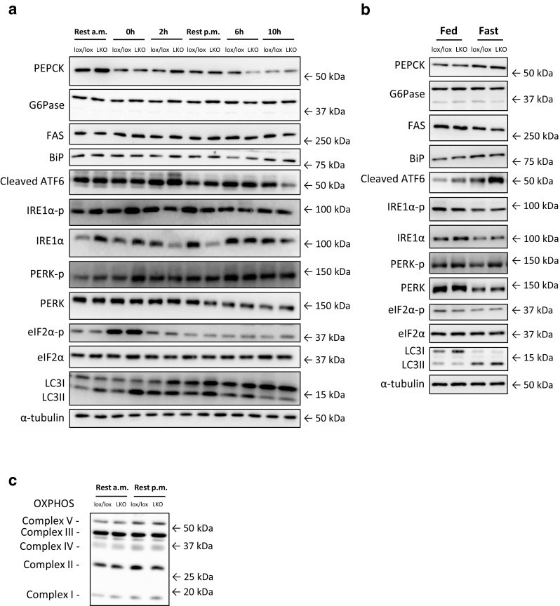
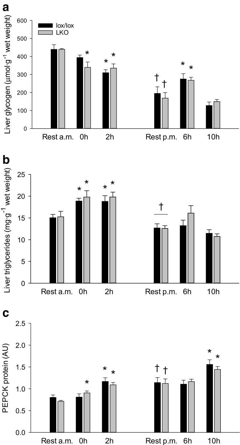
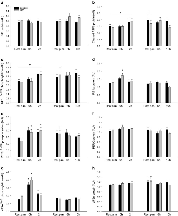
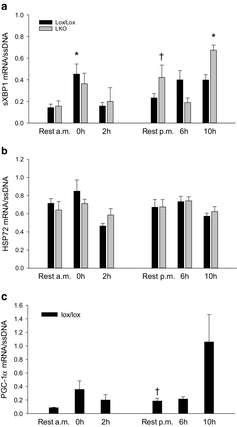
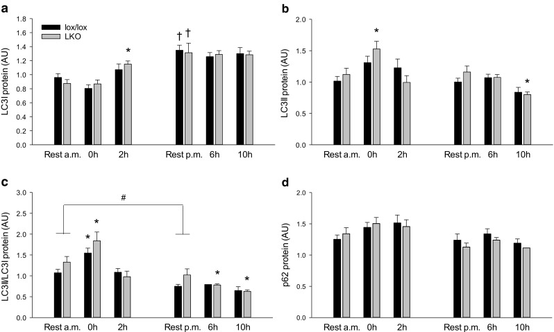
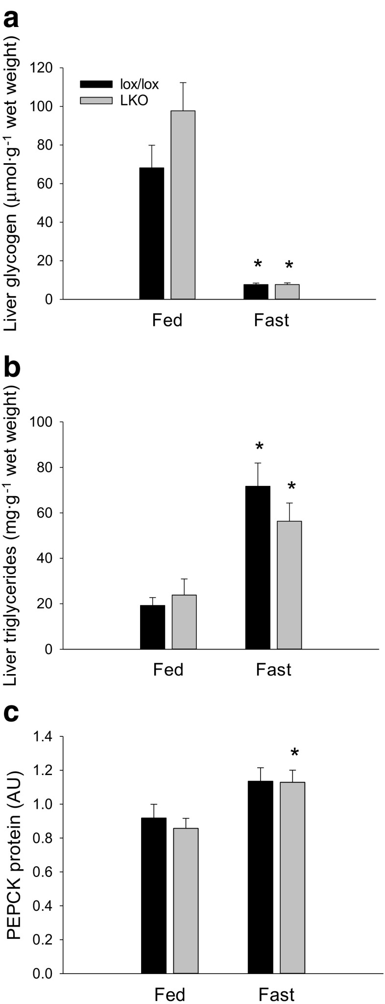
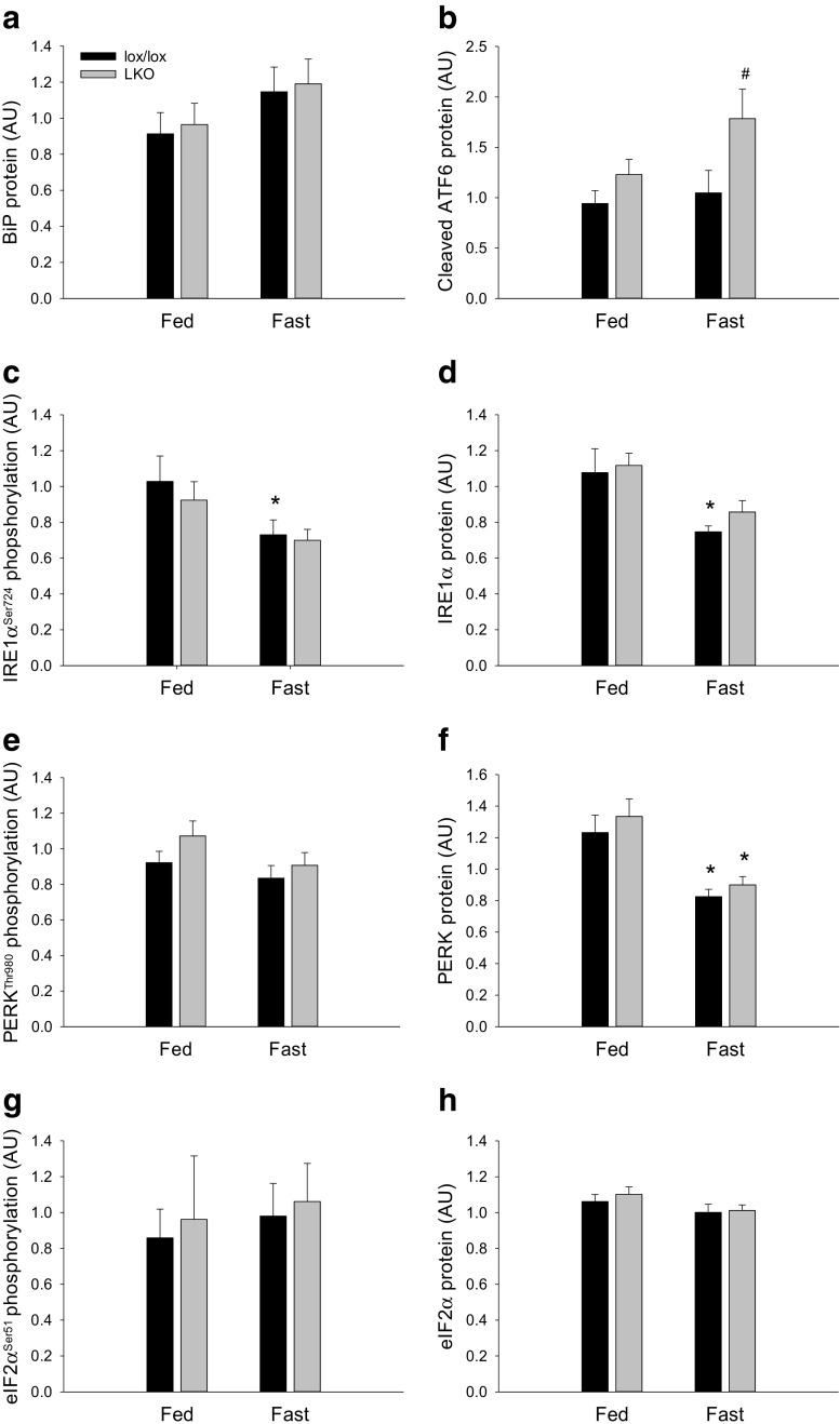
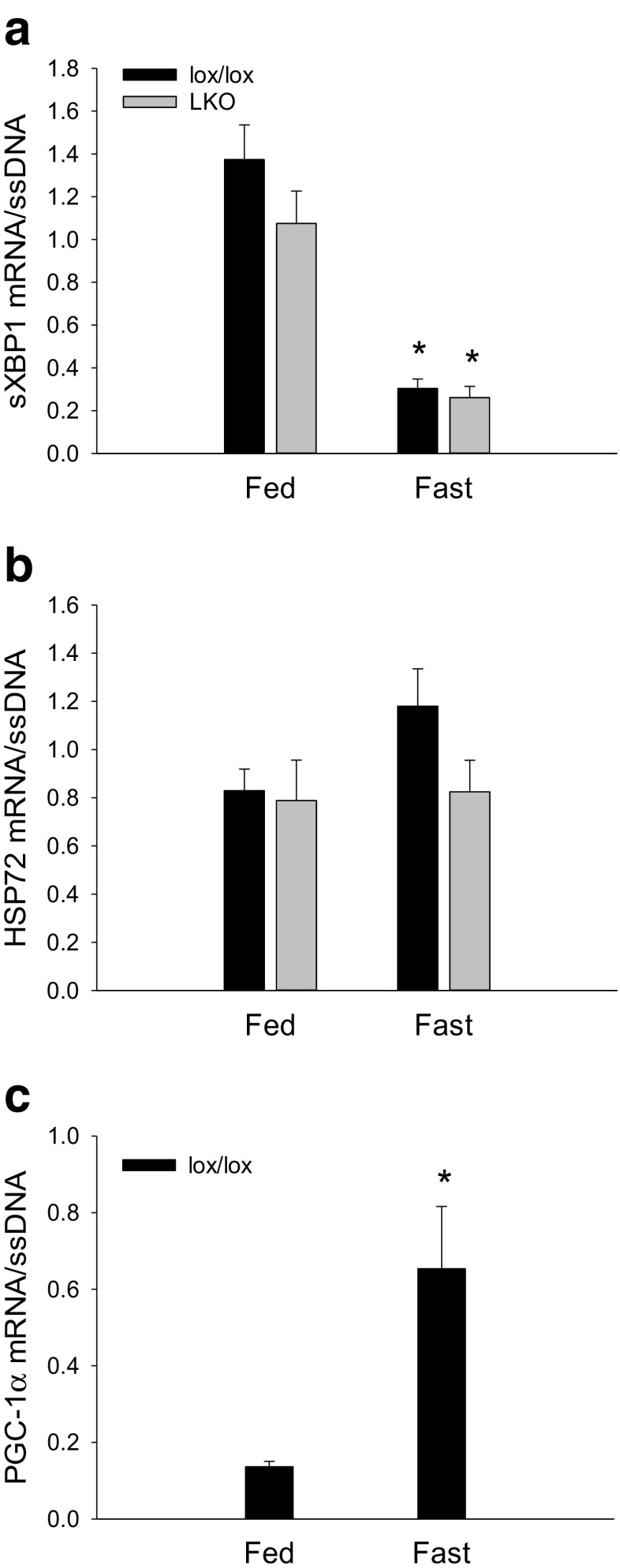
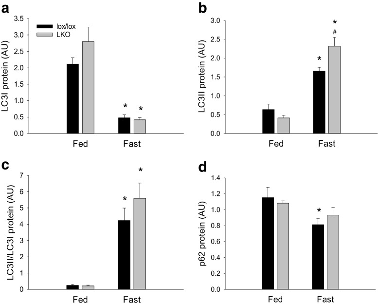
The aim of the present study was to test the hypothesis that PGC-1α is involved in the regulation of hepatic UPR and autophagy in response to both exercise and fasting in mice. Liver-specific PGC-1α knockout (LKO) mice and their floxed littermates (lox/lox) were used in two experimental parts. Liver and plasma were obtained from (1) fed and 18 h fasted mice and (2) immediately after, 2, 6, and 10 h after 1-h treadmill running as well as from resting mice, where one resting group was euthanized at time points corresponding to 0 and 2 h and another corresponding to 6 and 10 h of recovery. Hepatic eIF2α phosphorylation and sXBP1 mRNA content increased immediately after exercise and IRE1α phosphorylation as well as cleaved ATF6 protein content was higher 2 h into recovery than at rest in both genotypes. Fasting reduced hepatic IRE1α phosphorylation and protein content as well as PERK protein and sXBP1 mRNA content similarly in lox/lox and LKO mice. In addition, the hepatic LC3II/LC3I protein ratio increased immediately after exercise and with fasting in both genotypes, while fasting decreased p62 protein content in lox/lox mice. Liver-specific PGC-1α knockout did not affect these responses, but the LC3II/LC3I protein ratio was higher in LKO than lox/lox mice in both rest groups. In conclusion, the present study provides evidence for pathway-specific exercise-induced activation and fasting-induced downregulation of the UPR as well as exercise and fasting-induced regulation of autophagy in mouse liver. In addition, overall PGC-1α does not seem to be required for the fasting and exercise-induced regulation of UPR and autophagy, but may be involved in regulating basal hepatic autophagy.

Keywords: Exercise; Fasting; Liver; PGC-1α; UPR.


Conflict of interest statement

The authors declare that they have no conflicts of interest.

Figures

Fig. 1
Fig. 1
Representative western blots from the exercise (a) and fasting (b) study. PEPCK, phosphoenolpyruvate carboxykinase; G6Pase, glucose-6-phosphatase; FAS, fatty acid synthase; BiP, binding immunoglobulin protein; ATF6, activating transcription factor 6; IRE1α, inositol-requiring enzyme 1 alpha; PERK, protein kinase-like ER-kinase; eIF2α, eukaryotic translation initiation factor 2 alpha; LC3, microtubule-associated protein 1 light chain 3. In addition, representative western blots of OXPHOS in resting mice (c)
Fig. 2
Fig. 2
Hepatic glycogen (a), triglyceride (b), and PEPCK protein (c) content in liver-specific PGC-1α knockout (LKO) and littermate floxed (lox/lox) mice at rest and 0, 2, 6, and 10 h into recovery from 1-h treadmill running. Resting mice were either euthanized at time points corresponding to 0 and 2 h (rest a.m.) or 6 and 10 h (rest p.m.). Protein is given in arbitrary units (AU). Values are presented as means ± standard error (SE); n = 8–10. A single asterisk is significantly different from rest within given genotype, p < 0.05. A single dagger is significantly different from rest a.m. within given genotype, p < 0.05. Horizontal line indicates a main effect, p < 0.05
Fig. 3
Fig. 3
Hepatic BiP protein (a), cleaved ATF6 protein (b), IRE1αSer924 phosphorylation (c), IRE1α protein (d), PERKThr980 phosphorylation (e), PERK protein (f), eIF2αSer51 phosphorylation (g), and eIF2α protein (h) content in liver-specific PGC-1α knockout (LKO) and littermate floxed (lox/lox) mice at rest and 0, 2, 6, and 10 h into recovery from 1-h treadmill running. Resting mice were either euthanized at time points corresponding to 0 and 2 h (rest a.m.) or 6 and 10 h (rest p.m.). Protein content and phosphorylation are given in arbitrary units (AU). Values are presented as means ± standard error (SE); n = 8–10. A single asterisk is significantly different from rest within given genotype, p < 0.05. A single dagger is significantly different from rest a.m. within given genotype, p < 0.05. Horizontal line indicates a main effect, p < 0.05
Fig. 4
Fig. 4
Hepatic sXBP1 (a) and HSP72 (b) mRNA content in liver-specific PGC-1α knockout (LKO) and littermate floxed (lox/lox) mice and PGC-1α mRNA in lox/lox mice (c) at rest and 0, 2, 6, and 10 h into recovery from 1-h treadmill running. Resting mice were either euthanized at time points corresponding to 0 and 2 h (rest a.m.) or 6 and 10 h (rest p.m.). The target mRNA is normalized to single stranded (ss) DNA. Values are presented as means ± standard error (SE); n = 6–9. A single asterisk is significantly different from rest within given genotype, p < 0.05. A single dagger is significantly different from rest a.m. within given genotype, p < 0.05
Fig. 5
Fig. 5
Hepatic LC3I (a), LC3II (b), LC3II/LC3I protein ratio (c) and p62 protein (d) in liver-specific PGC-1α knockout (LKO) and littermate floxed (lox/lox) mice at rest and 0, 2, 6, and 10 h into recovery from 1-h treadmill running. Resting mice were either euthanized at time points corresponding to 0 and 2 h (rest a.m.) or 6 and 10 h (rest p.m.). Protein content is given in arbitrary units (AU). Values are presented as means ± standard error (SE); n = 8–10. A single asterisk is significantly different from rest within given genotype, p < 0.05. A single dagger is significantly different from rest a.m. within given genotype, p < 0.05. A single number sign is significantly different from lox/lox within given group, p < 0.05. Horizontal line indicates a main effect, p < 0.05
Fig. 6
Fig. 6
Hepatic glycogen (a), triglyceride (b), and PEPCK protein (c) content in fed (Fed) and 18 h fasted (Fast) liver-specific PGC-1α knockout (LKO), and littermate floxed (lox/lox) mice. Protein is given in arbitrary units (AU). Values are presented as means ± standard error (SE); n = 7–8. A single asterisk is significantly different from Fed within given genotype, p < 0.05
Fig. 7
Fig. 7
Hepatic BiP protein (a), cleaved ATF6 protein (b), IRE1αSer724 phosphorylation (c), IRE1α protein (d), PERKThr980 phosphorylation (e), PERK protein (f), eIF2αSer51 phosphorylation (g), and eIF2α protein (h) content in fed (Fed) and 18 h fasted (Fast) liver-specific PGC-1α knockout (LKO) and littermate floxed (lox/lox) mice. Protein is given in arbitrary units (AU). Values are presented as means ± standard error (SE); n = 7–8. A single asterisk is significantly different from Fed within given genotype, p < 0.05. A single number sign is significantly different from lox/lox within given group, p < 0.05
Fig. 8
Fig. 8
Hepatic sXBP1 (a) and HSP72 (b) mRNA content in fed (Fed) and 18 h fasted (Fast) liver-specific PGC-1α knockout (LKO) and littermate floxed (lox/lox) mice and PGC-1α mRNA in lox/lox mice (c). The target mRNA is normalized to single stranded (ss) DNA. Values are presented as means ± standard error (SE); n = 6–8. A single asterisk is significantly different from Fed within given genotype, p < 0.05
Fig. 9
Fig. 9
Hepatic LC3I protein (a), LC3II protein (b), LC3II/LC3I (c) and p62 protein (d) protein ratio in fed (Fed) and 18 h fasted (Fast) liver-specific PGC-1α knockout (LKO) and littermate floxed (lox/lox) mice. Protein content is given in arbitrary units (AU). Values are presented as means ± standard error (SE); n = 7–8. A single asterisk is significantly different from Fed within given genotype, p < 0.05. A single number sign significantly different from lox/lox within given group, p < 0.05

References

    1. Banzet S, Koulmann N, Simler N, Sanchez H, Chapot R, Serrurier B, Peinnequin A, Bigard X. Control of gluconeogenic genes during intense/prolonged exercise: hormone-independent effect of muscle-derived IL-6 on hepatic tissue and PEPCK mRNA. J Appl Physiol. 2009;107:1830–1839. doi: 10.1152/japplphysiol.00739.2009. - DOI - PubMed
    1. Bertholdt L, Gudiksen A, Schwartz CL, Knudsen JG, Pilegaard H. Lack of skeletal muscle IL-6 influences hepatic glucose metabolism in mice during prolonged exercise. Am J Phys Regul Integr Comp Phys. 2017;312:R626–R636. - PubMed
    1. Bertolotti A, Zhang Y, Hendershot LM, Harding HP, Ron D. Dynamic interaction of BiP and ER stress transducers in the unfolded-protein response. Nat Cell Biol. 2000;2:326–332. doi: 10.1038/35014014. - DOI - PubMed
    1. Birk JB, Wojtaszewski JF. Predominant alpha2/beta2/gamma3 AMPK activation during exercise in human skeletal muscle. J Physiol. 2006;577:1021–1032. doi: 10.1113/jphysiol.2006.120972. - DOI - PMC - PubMed
    1. Brandt N, Dethlefsen MM, Bangsbo J, Pilegaard H. PGC-1alpha and exercise intensity dependent adaptations in mouse skeletal muscle. PLoS One. 2017;12:e0185993. doi: 10.1371/journal.pone.0185993. - DOI - PMC - PubMed

Publication types

MeSH terms

Substances

LinkOut - more resources