. 2018 Jul 5;103(1):144-153.
doi: 10.1016/j.ajhg.2018.06.001. Epub 2018 Jun 28.

De Novo Truncating Mutations in WASF1 Cause Intellectual Disability with Seizures

Collaborators, Affiliations

De Novo Truncating Mutations in WASF1 Cause Intellectual Disability with Seizures

Yoko Ito et al. Am J Hum Genet. .

Abstract

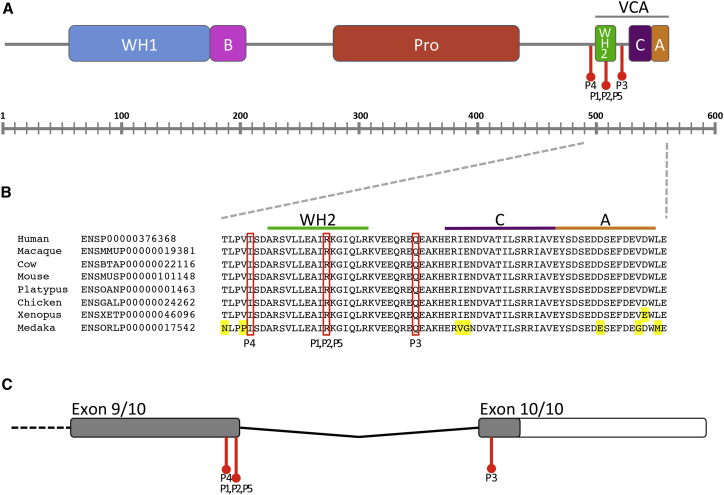
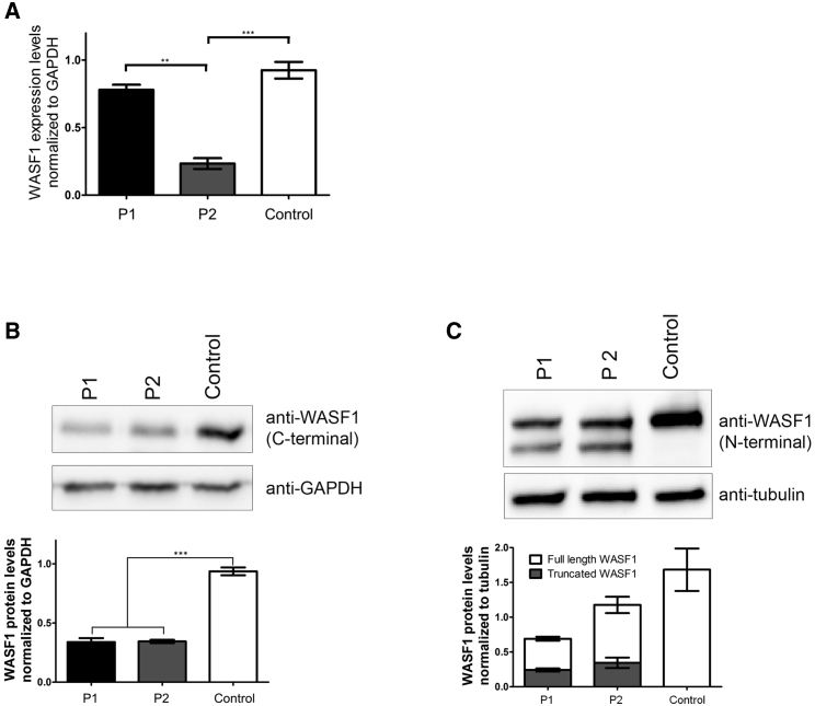
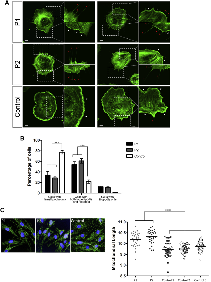
Next-generation sequencing has been invaluable in the elucidation of the genetic etiology of many subtypes of intellectual disability in recent years. Here, using exome sequencing and whole-genome sequencing, we identified three de novo truncating mutations in WAS protein family member 1 (WASF1) in five unrelated individuals with moderate to profound intellectual disability with autistic features and seizures. WASF1, also known as WAVE1, is part of the WAVE complex and acts as a mediator between Rac-GTPase and actin to induce actin polymerization. The three mutations connected by Matchmaker Exchange were c.1516C>T (p.Arg506Ter), which occurs in three unrelated individuals, c.1558C>T (p.Gln520Ter), and c.1482delinsGCCAGG (p.Ile494MetfsTer23). All three variants are predicted to partially or fully disrupt the C-terminal actin-binding WCA domain. Functional studies using fibroblast cells from two affected individuals with the c.1516C>T mutation showed a truncated WASF1 and a defect in actin remodeling. This study provides evidence that de novo heterozygous mutations in WASF1 cause a rare form of intellectual disability.

Keywords: WASF1; WAVE1 complex; actin cytoskeleton; autism; developmental delay; lamellipodia; neurodevelopmental disorder; recurrent de novo truncating mutations; seizures.


Figures

Figure 1
Figure 1
Schematic Diagrams Showing Structure of WASF1 and WASF1 (A) Schematic diagram showing full-length WASF1 (also known as WAVE1 [Ensembl: ENSP00000376368]). Variants in the five individuals (indicated in red) cluster around the WH2 domain (domain coordinates are from Stradal et al.14). P1, P2, and P5 have p.Arg506Ter, P3 has p.Gln520Ter, and P4 has p.Ile494MetfsTer23. Abbreviations are as follows: WH1, WASP homology 1 domain; B, basic domain; Pro, proline-rich region; WH2, WASP homology 2 domain (also known as the verprolin homology domain); C, cofilin homology domain; A, acidic domain; WCA, collective name for the WH2, C, and A domains. (B) Schematic diagram showing the amino acid sequence of part of WASF1. The WCA region of WASF1 is conserved throughout evolution. Yellow highlights residues that differ from the human protein sequence. (C) Schematic diagram showing the 3′ part of WASF1, including locations of the participants’ variants in red. The gray boxes represent the coding sequence, and the white box represents the 3′ UTR. The variant in P1, P2, and P5 is 6 bps from the end of exon 9 (the penultimate exon). The variant in P4 is 40 bps from the end of exon 9. The variant in P3 is within exon 10 (the final exon).
Figure 2
Figure 2
Amounts of WASF1 mRNA and WASF1 in Fibroblasts Derived from Affected Individuals with the c.1516C>T Variant (A) RT-qPCR shows variable amounts of WASF1 mRNA between primary fibroblasts derived from individuals P1 and P2 and healthy control fibroblasts. (B) Western blot analysis using an antibody with an epitope downstream of Arg506 showed that the amount of full-length WASF1 was approximately 50% lower in affected fibroblasts than in control fibroblasts. (C) Western blot analysis using an antibody with an epitope in the N-terminal region of WASF1 showed the presence of the full-length and truncated WASF1 in affected fibroblasts. The truncated WASF1 was not present in control fibroblasts. All experiments were performed with fibroblasts derived from three healthy control individuals. Western blots were performed in triplicate, and band intensity was quantified with Image Lab Software (Bio-Rad). Error bars indicate the range of measurement of triplicate samples.
Figure 3
Figure 3
Lamellipodia Formation and Mitochondrial Morphology in Fibroblasts Derived from Individuals with the c.1516C>T Variant (A) Primary fibroblasts were treated with PDGF for inducing the formation of lamellipodia. Visualization of the filamentous actin by phalloidin staining revealed the disruption of actin in the cell periphery of P1 and P2 fibroblasts. In the insets, lamellipodia and filopodia are marked by white arrowheads and red asterisks, respectively. Scale bars represent 10 μm. (B) Cells were categorized into three groups on the basis of the predominant actin phenotype present: cells displaying lamellipodia only, cells displaying a mixture of lamellipodia and filopodia, and cells displaying filopodia only. Quantification based on these three categories indicates that significantly fewer affected fibroblasts than control fibroblasts are able to form solely lamellipodia. (C) Confocal microscopic analysis of TOMM-20-immunostained mitochondria (in green) indicated that both affected fibroblasts have significantly elongated mitochondria. The nuclei were visualized by DAPI staining (in blue).

References

    1. Vissers L.E.L.M., Gilissen C., Veltman J.A. Genetic studies in intellectual disability and related disorders. Nat. Rev. Genet. 2016;17:9–18. - PubMed
    1. Martínez F., Caro-Llopis A., Roselló M., Oltra S., Mayo S., Monfort S., Orellana C. High diagnostic yield of syndromic intellectual disability by targeted next-generation sequencing. J. Med. Genet. 2017;54:87–92. - PubMed
    1. Veltman J.A., Brunner H.G. De novo mutations in human genetic disease. Nat. Rev. Genet. 2012;13:565–575. - PubMed
    1. Rauch A., Wieczorek D., Graf E., Wieland T., Endele S., Schwarzmayr T., Albrecht B., Bartholdi D., Beygo J., Di Donato N. Range of genetic mutations associated with severe non-syndromic sporadic intellectual disability: an exome sequencing study. Lancet. 2012;380:1674–1682. - PubMed
    1. Hamdan F.F., Srour M., Capo-Chichi J.-M., Daoud H., Nassif C., Patry L., Massicotte C., Ambalavanan A., Spiegelman D., Diallo O. De novo mutations in moderate or severe intellectual disability. PLoS Genet. 2014;10:e1004772. - PMC - PubMed

Publication types

Substances