. 2018 Jul 27;46(13):6401-6415.
doi: 10.1093/nar/gky529.

Structure of HIV TAR in complex with a Lab-Evolved RRM provides insight into duplex RNA recognition and synthesis of a constrained peptide that impairs transcription

Affiliations

Structure of HIV TAR in complex with a Lab-Evolved RRM provides insight into duplex RNA recognition and synthesis of a constrained peptide that impairs transcription

Ivan A Belashov et al. Nucleic Acids Res. .

Abstract

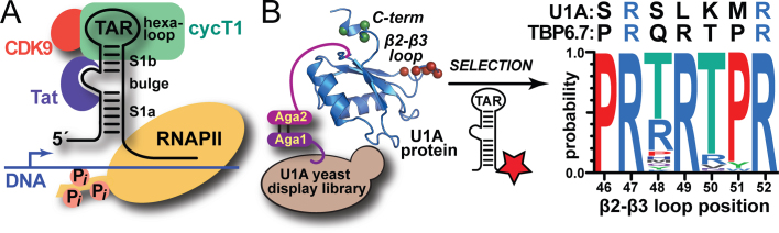
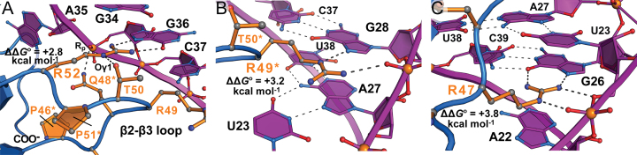
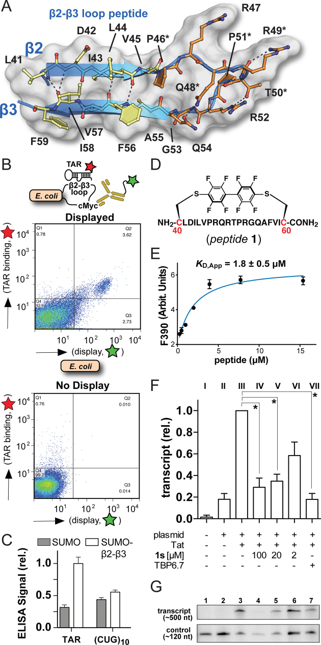
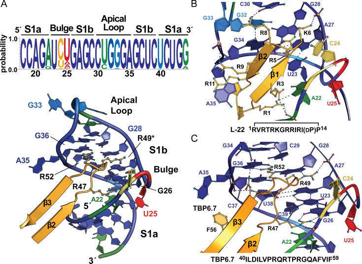
Natural and lab-evolved proteins often recognize their RNA partners with exquisite affinity. Structural analysis of such complexes can offer valuable insight into sequence-selective recognition that can be exploited to alter biological function. Here, we describe the structure of a lab-evolved RNA recognition motif (RRM) bound to the HIV-1 trans-activation response (TAR) RNA element at 1.80 Å-resolution. The complex reveals a trio of arginines in an evolved β2-β3 loop penetrating deeply into the major groove to read conserved guanines while simultaneously forming cation-π and salt-bridge contacts. The observation that the evolved RRM engages TAR within a double-stranded stem is atypical compared to most RRMs. Mutagenesis, thermodynamic analysis and molecular dynamics validate the atypical binding mode and quantify molecular contributions that support the exceptionally tight binding of the TAR-protein complex (KD,App of 2.5 ± 0.1 nM). These findings led to the hypothesis that the β2-β3 loop can function as a standalone TAR-recognition module. Indeed, short constrained peptides comprising the β2-β3 loop still bind TAR (KD,App of 1.8 ± 0.5 μM) and significantly weaken TAR-dependent transcription. Our results provide a detailed understanding of TAR molecular recognition and reveal that a lab-evolved protein can be reduced to a minimal RNA-binding peptide.


Figures

Figure 1.
Figure 1.
Cartoon depicting the dependence of HIV-1 transcription upon the viral TAR–Tat interaction and overview of the ‘semi design’ approach that led to TBP6.7 (TAR-Binding Protein 6.7). (A) The viral trans-activation response (TAR) element RNA comprises lower (S1a) and upper (S1b) stems. The positive transcription elongation factor b (p-TEFb) comprising cyclin T1 (green) and CDK9 (red) is recruited to TAR by the HIV-1 protein Tat (purple), which binds the central RNA bulge allowing cyclin T1 to interact with the apical loop. The bound complex stimulates host RNA polymerase II (yellow) by phosphorylation to produce full-length viral transcripts from proviral DNA [reviewed in (12)]. (B) A yeast-display approach was used to diversify putative RNA-binding amino acids in the β2–β3 loop and C-terminus of U1A RRM1 (RNA Recognition Motif 1, depicted as a blue ribbon); selection utilized labeled TAR (star) binding under increasingly stringent conditions (21). The resulting loop consensus sequence is shown (right) along with amino acids from U1A and TBP6.7—the tightest known TAR binder (21). R47 and R52 were unaltered to exploit their innate RNA binding potential.
Figure 2.
Figure 2.
Ribbon and schematic diagrams depicting the HIV-1 TAR–TBP6.7 complex of this investigation and parental U1hpII-U1A. (A) Global view of the co-crystal structure depicting the TBP6.7 RRM domain (blue) engaging TAR RNA (purple) in upper helical stem S1b. Arginines of the β2–β3 loop that provide the principal determinants of TAR binding are depicted as ball-and-stick models (orange); similar depictions are provided for conserved RRM amino acids known as RNP2 (Y13) and RNP1 (R52, Q54 and F56). (B) Global view of the structure in A rotated +90°, providing a view looking through the apical loop and down the helical axis. The TBP6.7 β2–β3 loop penetrates deeply into the TAR major groove. (C) Schematic diagram depicting interactions between TBP6.7 and TAR based on the co-crystal structure. Henceforth asterisks (*) indicate lab-evolved TBP6.7 residues depicted in Figure 1B. (D) Close-up of the TAR Uri23•Ade27-Uri38 major-groove base triple and the central bulge that interrupts stems S1a and S1b. Dashed lines joining ball-and-stick models represent putative hydrogen bonds unless noted otherwise. (E) Close-up view of the apical hexaloop and interface with the S1b closing base pair. (F) Global view of the U1hpII-U1A complex (23) oriented and colored as in A. U1A binds U1hpII primarily within the single-stranded region of the upper loop.
Figure 3.
Figure 3.
Close-up views of key interactions between the evolved β2–β3 hairpin loop of TBP6.7 and HIV-1 TAR based on the co-crystal structure. ΔΔG° values from ITC analysis of R-to-A mutations are taken from Supplementary Table S1. (A) R52 forms two hydrogen bonds to the Hoogsteen edge of Gua36; for clarity, some evolved amino acids in the β2–β3 loop are omitted. (B) R49* forms a hydrogen bond with N7 of Gua28 and a salt-bridge to its non-bridging phosphate oxygen. (C) R47 forms two hydrogen bonds with the Hoogsteen edge of Gua26, as well as hydrogen bond and salt-bridge interactions to the Uri23 phosphate. Cation–π interactions and buried surface areas for each arginine are described in Supplementary Figure S5.
Figure 4.
Figure 4.
Cartoon depiction of the β2–β3-loop peptide supersecondary structure and retention of TAR binding by β2–β3-loop peptides outside the context of TBP6.7. (A) Diagram illustrating hydrogen bonds within the close-packed β-strand core (transparent surface and yellow amino acids) and loop (orange amino acids) of the TAR–TBP6.7 complex. Key TAR-binding arginines are shown, as well as wild-type and evolved amino acids that contribute peptide stability. (B) Fluorescence activated cell-sorting analysis of TAR binding. (Upper) A fusion protein expressed on the E. coli surface harbors the TBP6.7 β2–β3-loop peptide from A. (Lower) Display and TAR binding control using only E. coli. (C) ELISA analysis of TAR binding by SUMO (control) and a SUMO-β2–β3-loop fusion protein harboring the sequence from A; off target (CUG)10 RNA is used as a binding control. (D) Schematic drawing of conformationally-constrained peptide1 derived from the sequence in A, except cysteine termini were added (red) for conjugation to polyfluorinated biphenyl. (E) Titration of peptide1 into 2-aminopurine-labeled TAR produces changes in fluorescence emission at 390 nm. Filled circles represent the experimental data resulting from three independent measurements. The smooth curve shows the fit of a one-site-binding model, which gave the apparent KD with standard deviation shown. (F) Graphed densitometry data of HIV-1 TAR–Tat-dependent transcription in HeLa nuclear lysate and inhibition by peptide1s or TBP6.7. P values from t tests are: 100 μM, P = 0.040; 20 μM, P = 0.028; 2 μM, P = 0.055 and 10 μM TBP6.7, P = 0.013. *P < 0.05 is significant and P > 0.05 is not significant. (G) Representative densitometry signals for TAR–Tat-dependent transcription in HeLa nuclear extract, and suppression of transcription by peptide1s or TBP6.7. For C and F, data are plotted as the mean of three separate experiments with corresponding standard errors of the mean. An intact gel used in F and G is provided (Supplementary Figure S9).
Figure 5.
Figure 5.
HIV-1 TAR conservation and comparison of the TAR–TBP6.7 complex to a known antiviral cyclic peptide. (A) (Upper) Sequence conservation for representative circulating forms of HIV-1 depicted as a web-logo diagram. Blue represents greatest conservation and red indicates poor conservation. (Lower) Cartoon diagram of the TAR–TBP6.7 complex with web-logo sequence conservation heat mapped onto the RNA. (B) The TAR–L-22 complex (PDB entry 2kdq) (17) reveals interactions distributed throughout the RNA-peptide interface, which yields a shape-complementarity score (Sc) of 0.60, indicating a high degree of interlocking surface. (C) The TAR–TBP6.7 complex shows a clustered trio of arginines that recognizes three highly conserved guanosine nucleotides by hydrogen bond, salt-bridge and cation-π contacts (Figure 3 and Supplementary Figure S5). Here, the Sc score is 0.79 for the co-crystal structure and 0.76 for the isolated β2–β3 loop peptide shown. These scores are comparable to the nearly ideal Sc score of 0.84 measured for the U1A–hpII interface. The Uri23•Ade27-U38 triple is intact in the presence of a canonical Cyt30–Gua34 pair located in the apical loop.
Figure 6.
Figure 6.
Representative atypical RRMs that recognize the RNA major groove. (A) Ribbon diagram of the human RBMY-CA/CAA pentaloop complex. Although this naturally occurring RRM uses classical single-stranded RNA recognition by RNP1 and RNP2 amino acids, its β2–β3-loop residues engage in modest double-stranded RNA readout (PDB entry 2fyi) (86). RNP residues are depicted as ball-and-stick models colored similarly to those of Figure 2A,F; idiosyncratic protein residues that recognize duplex RNA are colored lime green. (B) Ribbon diagram of the Bacillus subtilis YxiN protein in complex with 23S rRNA (PDB entry 3moj) (87). Salt bridges to the backbone and hydrophobic contacts form an array of complementary interactions between the RRM and the three-way helical junction major groove. RNP and β2–β3-loop residues are not used in RNA binding. (C) Ribbon diagram of the p65 C-terminal RRM (p65-C1ΔL2:S4) in complex with telomerase RNA stem IV (PDB entry 4erd) (88). An unusually long β2–β3 loop was truncated for structural studies but is dispensable for RNA binding. The atypical mode of double-stranded RNA binding utilizes a C-terminal α-helical extension that interacts with the major groove, along with idiosyncratic amino acids contributed from strands β2 and β3. Single- and double-stranded RNA recognition occurs without use of RNP amino acids.

Similar articles

Cited by

References

    1. Fauci A., Dieffenbach C.. Thirty Years of HIV and AIDS: future challenges and opportunities. Ann. Intern. Med. 2011; 154:766–771. - PubMed
    1. HIV/AIDS, J.U.N.P.o. 2016; Geneva: www.unaids.org/sites/default/files/media_asset/global-AIDS-update-2016_e....
    1. Cary D.C., Peterlin B.M.. Targeting the latent reservoir to achieve functional HIV cure. F1000Res. 2016; 5:F1000. - PMC - PubMed
    1. Harrich D., Ulich C., Gaynor R.B.. A critical role for the TAR element in promoting efficient human immunodeficiency virus type 1 reverse transcription. J. Virol. 1996; 70:4017–4027. - PMC - PubMed
    1. Huthoff H., Berkhout B.. Mutations in the TAR hairpin affect the equilibrium between alternative conformations of the HIV-1 leader RNA. Nucleic Acids Res. 2001; 29:2594–2600. - PMC - PubMed

Publication types

MeSH terms