. 2018 Aug 1;141(8):2392-2405.
doi: 10.1093/brain/awy171.

De novo variants in GABRA2 and GABRA5 alter receptor function and contribute to early-onset epilepsy

Affiliations

De novo variants in GABRA2 and GABRA5 alter receptor function and contribute to early-onset epilepsy

Kameryn M Butler et al. Brain. .

Abstract

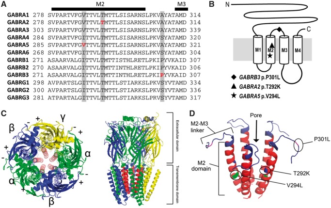
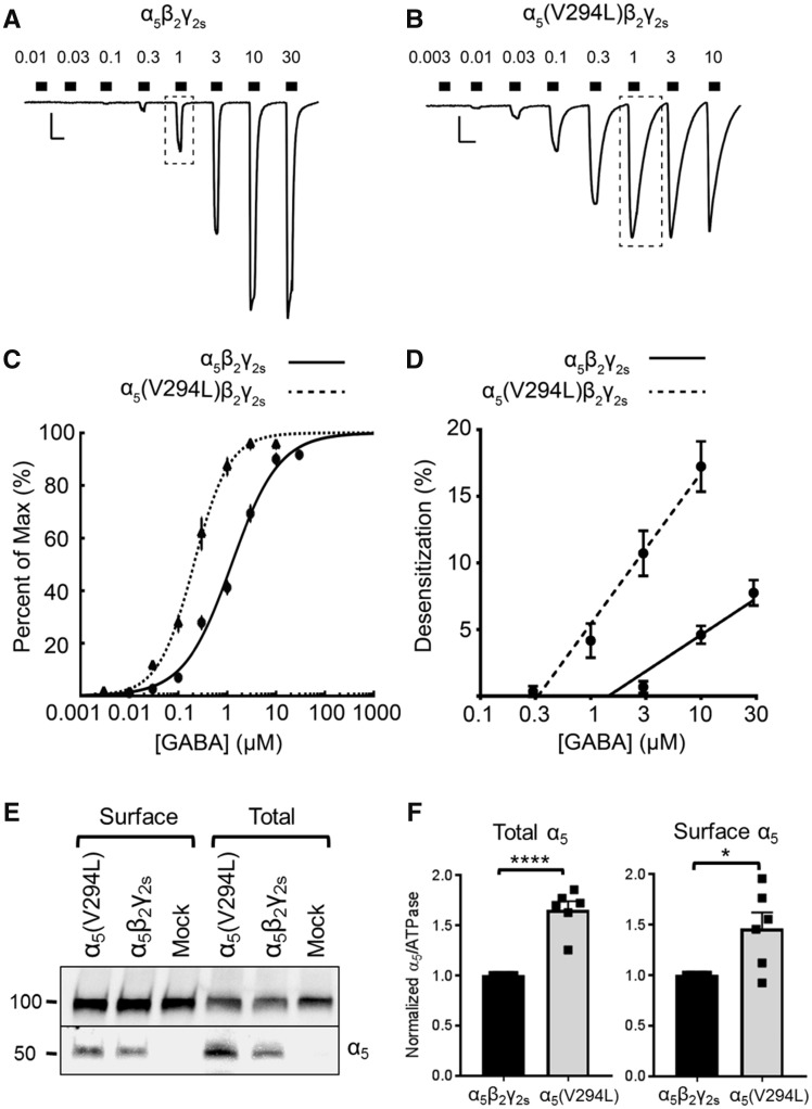
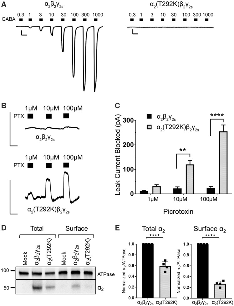
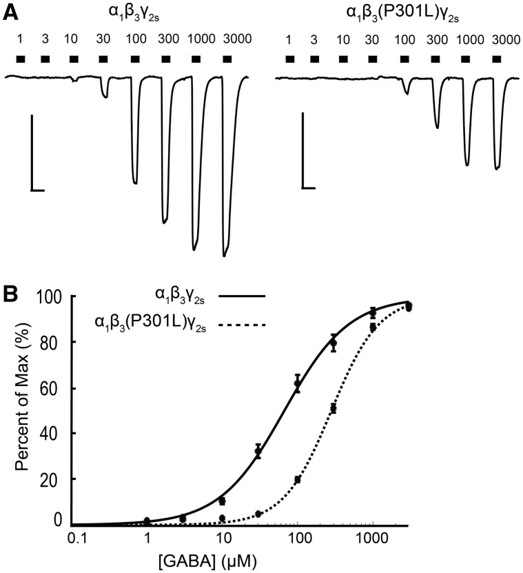
GABAA receptors are ligand-gated anion channels that are important regulators of neuronal inhibition. Mutations in several genes encoding receptor subunits have been identified in patients with various types of epilepsy, ranging from mild febrile seizures to severe epileptic encephalopathy. Using whole-genome sequencing, we identified a novel de novo missense variant in GABRA5 (c.880G > C, p.V294L) in a patient with severe early-onset epilepsy and developmental delay. Targeted resequencing of 279 additional epilepsy patients identified 19 rare variants from nine GABAA receptor genes, including a novel de novo missense variant in GABRA2 (c.875C > A, p.T292K) and a recurrent missense variant in GABRB3 (c.902C > T, p.P301L). Patients with the GABRA2 and GABRB3 variants also presented with severe epilepsy and developmental delay. We evaluated the effects of the GABRA5, GABRA2 and GABRB3 missense variants on receptor function using whole-cell patch-clamp recordings from human embryonic kidney 293T cells expressing appropriate α, β and γ subunits. The GABRA5 p.V294L variant produced receptors that were 10-times more sensitive to GABA but had reduced maximal GABA-evoked current due to increased receptor desensitization. The GABRA2 p.T292K variant reduced channel expression and produced mutant channels that were tonically open, even in the absence of GABA. Receptors containing the GABRB3 p.P301L variant were less sensitive to GABA and produced less GABA-evoked current. These results provide the first functional evidence that de novo variants in the GABRA5 and GABRA2 genes contribute to early-onset epilepsy and developmental delay, and demonstrate that epilepsy can result from reduced neuronal inhibition via a wide range of alterations in GABAA receptor function.


Figures

Figure 1
Figure 1
Location of epilepsy variants in the GABAA receptor subunits. (A) Alignment of human α, β, and γ subunits. The location of the α5(V294L), α2(T292K), and β3(P301L) variants are highlighted in grey and the specific amino acids affected are bolded in red. Secondary structures (M2 and M3 transmembrane domains) are also shown above the alignment. The following protein sequences were used to make the alignment: GABRA1, NP_000797; GABRA2, NP_000798; GABRA3, NP_000799; GABRA4, NP_000800; GABRA5, NP_000801; GABRA6, NP_000802; GABRB1, NP_000803; GABRB2, NP_000804; GABRB3, NP_000805; GABRG1, NP_775807; GABRG2, NP_000807; GABRG3, NP_150092. (B) Schematic representation of a single GABAA receptor subunit with the approximate locations of the three missense variants shown. (C) Top-down and side view of an assembled GABAA receptor containing two α (green), two β (blue), and one γ (yellow) subunit. The M2 domain of each subunit has been coloured red to show the location of the channel pore. (D) 3D representation of the five M2 domains of the assembled GABAA receptor pore with the three patient variants indicated.
Figure 2
Figure 2
Distribution of variants from the gnomAD database across the three GABAA receptor proteins. In each lollipop diagram, the ligand binding domain is shown in green, the transmembrane domain, which includes all four transmembrane segments, is shown in red, and the unstructured regions are shown in grey. The numbers beneath each figure refer to the amino acid number. Missense (blue) and synonymous (grey) variants observed in the gnomAD database are shown. Red lollipops represent the candidate variants identified in this study. (A) Lollipop diagram for α5 (encoded by GABRA5) showing the location of the p.V294L variant in the transmembrane region of the receptor. Protein domain annotation is based on UniProt accession P31644. (B) Lollipop diagram for the α2 receptor showing the location of the p.T292K variant. Based on UniProt accession P47869. (C) Lollipop diagram of the β3 receptor showing the location of p.P301L (red) as well as the locations of previously reported pathogenic GABRB3 variants (gold). Based on UniProt accession P28472.
Figure 3
Figure 3
Increased GABA apparent-affinity and increased desensitization of α5(V294L)β2γ2s receptors. (A and B) Example traces of GABA concentration-response assays for (A) α5β2γ2s receptors (0.01–30 μM) and (B) α5(V294L)β2γ2s receptors (0.003–10 μM) expressed in HEK293T cells. Scale bars: horizontal = 5 s, vertical = 500 pA. Dotted boxes highlight the 1 μM GABA response, which is larger and more desensitized in mutant receptors. (C) GABA concentration-response curves for α5β2γ2s (solid line, n = 18 cells) and α5(V294L)β2γ2s (dotted line, n = 22 cells) receptors. Points are mean ± SEM and error bars are not shown where bars are smaller than points. Lines are a representative fit based on the average GABA concentration responses. (D) Average desensitization of peak currents from α5β2γ2s (solid line) and α5(V294L)β2γ2s receptors (dotted line). Linear regressions to calculate desensitization are: y = 5.508x + 0.909 (α5β2γ2s) and y = 9.584x – 6.277 [α5(V294L)], where y is the per cent of desensitization, and x is the log[GABA] in micromolar. Points represent mean ± SEM. (E) Total and cell surface protein lysates were analysed by SDS-PAGE and blotted by anti-α5 and anti-ATPase antibodies. Experiments were performed in triplicate on protein samples from two separate transfections. A representative western blot is shown. (F) Band intensities of α5 protein were normalized to the ATPase signal. Bars represent mean ± SEM. An unpaired t-test was used to determine significance. *P ≤ 0.05, ****P < 0.0001.
Figure 4
Figure 4
α2(T292K)β2γ2s receptors are predominantly open and produce leak current that can be blocked by picrotoxin. (A) Example leak-subtracted trace of GABA concentration-response assays (0.3–1000 μM) for α2β2γ2s and α2(T292K)β2γ2s receptors expressed in HEK293T cells. Traces are aligned for easier visualization, although the mutant example starts at a greater baseline leak current. Scale bars: horizontal = 5 s, vertical = 500 pA. (B) Picrotoxin (PTX) blocked tonic leak current of α2(T292K)β2γ2s receptors in the absence of GABA, while wild-type α2β2γ2s receptors showed little block [wild-type α2: n = 4 cells; α2(T292K): n = 10 cells]. Scale bars: horizontal = 5 s, vertical = 300 pA. (C) Quantification of the leak current (pA) suppressed by picrotoxin. Picrotoxin block was significantly larger for mutant receptors at concentrations 10 μM (P = 0.0017) and 100 μM (P < 0.0001) (two-way repeated-measures ANOVA, Sidak post hoc test). Bars represent mean ± SEM. **P ≤ 0.01, ****P < 0.0001, respectively. (D) Total and cell surface protein lysates were blotted with anti-α2 and anti-ATPase antibodies. Experiments were performed in duplicate on protein samples from two separate transfections. A representative western blot is shown. (E) Band intensities of α2 protein were normalized to the ATPase signal. Bars represent mean ± SEM. An unpaired t-test was used to determine significance. ****P < 0.0001.
Figure 5
Figure 5
α1β3(P301L)γ2s receptors are less sensitive to GABA activation. (A) Example trace of GABA concentration-response assays (1–3000 μM) for α1β3γ2s and α1β3(P301L)γ2s receptors expressed in HEK293T cells. Scale bars: horizontal = 5 s, vertical = 500 pA. (B) GABA concentration-response curves for α1β3γ2s (solid line, n = 21 cells) and α1β3(P301L)γ2s (dotted line, n = 20 cells) receptors. Points are mean ± SEM and error bars are not shown where bars are smaller than points. The drawn line is a representative fit based on the average GABA concentration responses.

Comment in

References

    1. Allen AS, Berkovic SF, Cossette P, Delanty N, Dlugos D, Eichler EE et al. . De novo mutations in epileptic encephalopathies. Nature 2013; 501: 217–21. - PMC - PubMed
    1. Baulac S, Huberfeld G, Gourfinkel-An I, Mitropoulou G, Beranger A, Prud’homme JF et al. . First genetic evidence of GABA(A) receptor dysfunction in epilepsy: a mutation in the gamma2-subunit gene. Nat Genet 2001; 28: 46–8. - PubMed
    1. Ben-Ari Y. Seizures beget seizures: the quest for GABA as a key player. Crit Rev Neurobiol 2006; 18: 135–44. - PubMed
    1. Bennett CA, Petrovski S, Oliver KL, Berkovic SF. ExACtly zero or once: a clinically helpful guide to assessing genetic variants in mild epilepsies. Neurol Genet 2017; 3: e163. - PMC - PubMed
    1. Bonin RP, Martin LJ, MacDonald JF, Orser BA. Alpha5GABAA receptors regulate the intrinsic excitability of mouse hippocampal pyramidal neurons. J Neurophysiol 2007; 98: 2244–54. - PubMed

Publication types