Intraoperative magnetic resonance imaging for neurosurgery - An anaesthesiologist's challenge
- PMID: 29962521
- PMCID: PMC6004746
- DOI: 10.4103/ija.IJA_29_18
Intraoperative magnetic resonance imaging for neurosurgery - An anaesthesiologist's challenge
Abstract
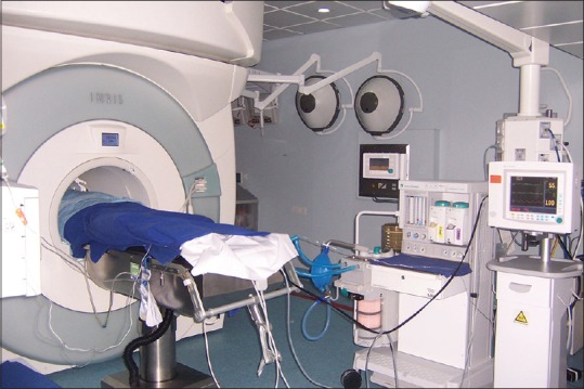
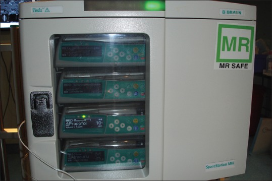
Intraoperative magnetic resonance imaging (MRI)-guided neurosurgery has gained popularity over the years globally. These surgeries require a dedicated operating room and MRI-compatible anaesthesia equipment. The anaesthesiologist providing care in this setup needs to be experienced and vigilant to ensure patient safety. Strict adherence to MRI safety checklists and regular personnel training would avoid potential accidents and life-threatening emergencies. Teamwork, good communication, preprocedure planning, and familiarity with the surroundings are very important for safe care and good outcomes. We performed a literature search in Google Scholar, PubMed and Cochrane databases for original and reviewed articles for the origins, development and applications of intraoperative MRI in neurosurgical procedures. Much of the research has emphasised on the surgical indications than the anaesthetic challenges faced during intraoperative MRI guided surgery. The purpose of this review is to discuss the anaesthetic concerns specific to this unique environment.
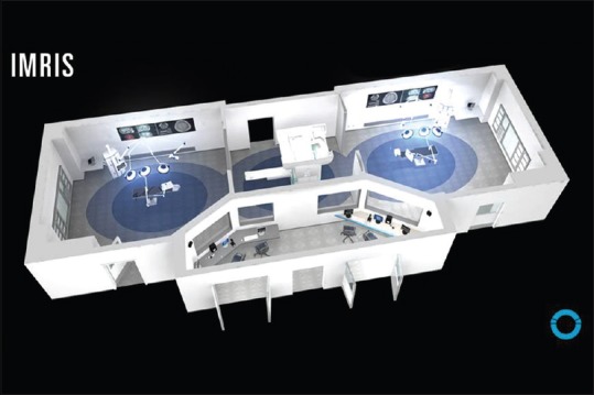
Keywords: Anaesthesia; IMRIS; MRI safe anaesthesia equipment; MRI safety checklist; awake craniotomy; challenge; electrical noise; intraoperative MRI; neurosurgery.
Conflict of interest statement
There are no conflicts of interest.
Figures
Similar articles
-
Magnetic resonance imaging-guided neurosurgery in the magnetic fringe fields: the next step in neuronavigation.Neurosurgery. 2000 Mar;46(3):643-53; discussion 653-4. doi: 10.1097/00006123-200003000-00023. Neurosurgery. 2000. PMID: 10719861
-
Anesthetic management and human factors in the intraoperative MRI environment.Curr Opin Anaesthesiol. 2016 Oct;29(5):563-7. doi: 10.1097/ACO.0000000000000366. Curr Opin Anaesthesiol. 2016. PMID: 27285726 Review.
-
Intraoperative magnetic resonance imaging-guided neurosurgery at 3-T.Neurosurgery. 2006 Apr;58(4 Suppl 2):ONS-338-45; discussion ONS-345-6. doi: 10.1227/01.NEU.0000205284.04252.87. Neurosurgery. 2006. PMID: 16582658
-
Anaesthesiologist's Approach to Awake Craniotomy.Turk J Anaesthesiol Reanim. 2018 Aug;46(4):250-256. doi: 10.5152/TJAR.2018.56255. Epub 2018 Aug 1. Turk J Anaesthesiol Reanim. 2018. PMID: 30140530 Free PMC article. Review.
-
Intraoperative magnetic resonance imaging with the magnetom open scanner: concepts, neurosurgical indications, and procedures: a preliminary report.Neurosurgery. 1998 Oct;43(4):739-47; discussion 747-8. doi: 10.1097/00006123-199810000-00005. Neurosurgery. 1998. PMID: 9766299
Cited by
-
Intraoperative Magnetic Resonance Imaging (iMRI) mishaps - Troubleshooting an unsafe object attached to the scanner.Indian J Anaesth. 2021 Mar;65(Suppl 1):S50-S51. doi: 10.4103/ija.IJA_870_20. Epub 2021 Mar 20. Indian J Anaesth. 2021. PMID: 33814591 Free PMC article. No abstract available.
-
Utilization of Portable Brain Magnetic Resonance Imaging in an Acute Care Setting.Cureus. 2022 Dec 28;14(12):e33067. doi: 10.7759/cureus.33067. eCollection 2022 Dec. Cureus. 2022. PMID: 36726935 Free PMC article.
-
Nerve autofluorescence in near-ultraviolet light markedly enhances nerve visualization in vivo.Surg Endosc. 2022 Mar;36(3):1999-2005. doi: 10.1007/s00464-021-08484-0. Epub 2021 Apr 9. Surg Endosc. 2022. PMID: 33835251
-
Supervised Machine Learning Methods and Hyperspectral Imaging Techniques Jointly Applied for Brain Cancer Classification.Sensors (Basel). 2021 May 31;21(11):3827. doi: 10.3390/s21113827. Sensors (Basel). 2021. PMID: 34073145 Free PMC article.
-
Deep Learning-Based Framework for In Vivo Identification of Glioblastoma Tumor using Hyperspectral Images of Human Brain.Sensors (Basel). 2019 Feb 22;19(4):920. doi: 10.3390/s19040920. Sensors (Basel). 2019. PMID: 30813245 Free PMC article.
References
-
- Bergese SD, Puente EG. Anaesthesia in the intraoperative MRI environment. Neurosurg Clin N Am. 2009;20:155–62. - PubMed
-
- Black PM, Moriarty T, Alexander E, Steig P, Woodard EJ, Langham Gleason P, et al. The development and implementation of intraoperative MRI and its neurosurgical applications. Neurosurgery. 1997;41:831–42. - PubMed
-
- Seifert V. Intraoperative MRI in neurosurgery: Technical overkill or the future of brain surgery? Neurol India. 2003;51:329–32. - PubMed
-
- Kettenbach J, Kacher DK, Koskinen SK, Silvermann SG, Nabavi A, Gering D, et al. Interventional and intraoperative magnetic resonance imaging. Annu Rev Biomed Eng. 2000;02:661–90. - PubMed